
Page 70
May 24-25, 2018
London, UK
Vascular Surgery 2018
3
rd
Edition of World Congress & Exhibition on
Vascular Surgery
Journal of Vascular and Endovascular Therapy
ISSN: 2573-4482
A penile venous stripping has been effective for treating erectile
dysfunction(ED)since1986.Weconductaretrospectiveanalysisto
thosewho received the latest method of surgery on an ambulatory
basis. From 2009 to 2016, 452 patients had a diagnosis of veno-
occlusive dysfunction (VOD). Of these, 283 men underwent the
latest method of penile venous stripping. They were divided into
young (n=46, younger than 30 years) and older (n=237) group
respectively. The surgery begins with a circumferential incision
followed by identification and management of the deep dorsal
veins (DDV) 1.5~2.5 cm proximal to the retro coronal sulcus. It
was then thoroughly stripped and ligated with 6-0 nylon sutures
with a pull-through maneuver. The cavernosal veins (CVs) were
managed in a similar manner. The para-arterial veins (PAVs) were
only segmentally ligated. Amedian longitudinal pubic incisionwas
then made to relay the stripping of the DDVs and CVs proximally
to the infrapubic angle. Finally, the pubic and circumferential
wounds were fashioned. A postoperative cavernosography was
made immediately. The operative times were 4.1±0.7 and 4.0±0.6
hr. respectively. The follow-up period ranged 1.2~7.2 (5.3±1.2)
years. Differences in erectile function were significant between
the groups of young and older group in term of preoperative IIEF-
5 (n=33, 10.2±3.6 vs. n=212, 9.7±3.8) scores compared to either
one-year postoperative (n=46, 19.1±3.2 vs. n=237, 16.4±3.0) ones
or two years postoperative (21.3±1.7 vs. 18.2±3.2) respectively
(both p<0.003). Overall, 92.3% (261/283) of the patients
reported improvements. On the preoperative and postoperative
cavernosograms, it was unexceptionally enhanced from weaker
to stronger radiopacity by this penile venous stripping. This latest
method of penile venous stripping appears to be a viable option
which achieves favorable outcomes with negligible morbidity for
treating ED secondary to VOD.
Analysis of the latest method of penile venous stripping
surgery in patient with veno-occlusive dysfunction
Chun Kai Hsu
1
and
Geng Long Hsu
2, 3
1
Taipei Tzu Chi Hospital, Taiwan
2
Hsu’s Andrology, Taiwan
3
National Taiwan University, Taiwan
Chun Kai Hsu et al., J Vasc Endovasc Therapy 2018, Volume 3
DOI: 10.21767/2573-4482-C1-002
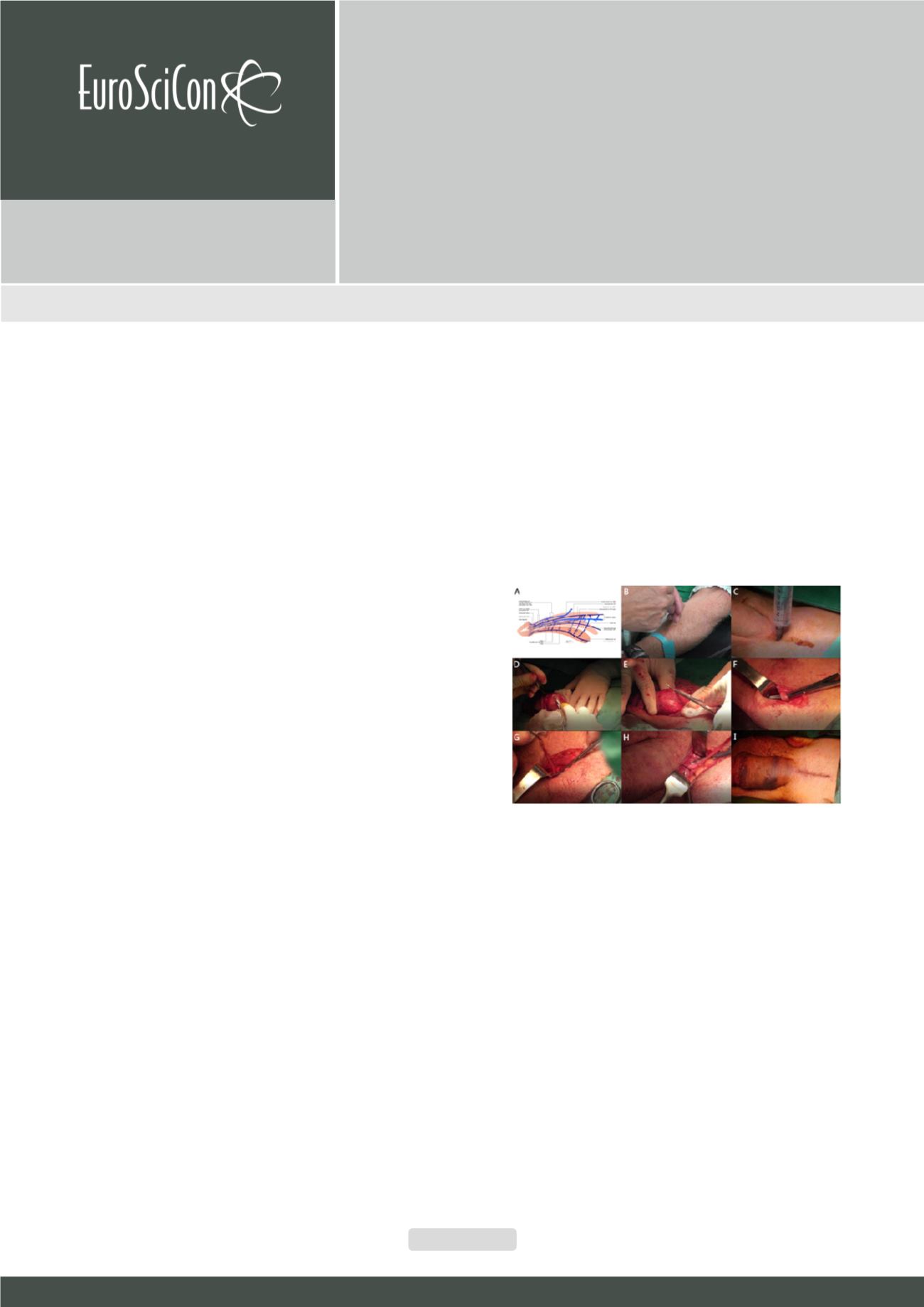
Figure 1:
Schematic illustration and blueprint and ongoing penile venous
stripping. (A) This is the blueprint for the latest method of penile venous
stripping. It entails the new insight into erection-related veins which require
being ligated at the tunical level to each emissary vein. In between the tunica
albuginea and Buck’s fascia, there is one deep dorsal vein (DDV), a couple
of cavernosal veins (CVs) and two pairs of para-arterial veins (PAVs), in con-
trast to conventional one – just one single DDV. DDV is consistently in the
median position and receives the blood of sinusoids of the glans penis and
the emissary veins from the corpora cavernosa and of the circumflex vein
from the corpus spongiosum. (B) Acupuncture is made on the acupoints of
Shou San Li (LI10)、Hegu (LI4) 、Quchi (LI11) and Waiguan (SJ5) . (C) Local
anesthesia is fulfilled via proximal dorsal nerve block、crural block and peipe-
nile infiltration. (D) The stripping surgery is initiated with a circumferential
incision followed by degloving those tissues superficial to the Colles’ fascia.
The visibility of the DDV can be enhanced by squeezing the corpora caverno-
sa. (E) Using a pull-through maneuver, opening on Buck’s fascia is made to
treat the emissary vein 5-6 times until the penile base. Likewise, the CVs are
managed. (F) A longitudinal pubic incision is made to relay the procedure.
(G) The DDV and CVs are treated respectively. (H) As a rule there are 6-9 and
5-8 big branches to DDV and CVs respectively. (I) In our experience, a total of
76–132 ligature sites are required to finish the penile venous stripping. Both
wounds are fashioned


