


 44 / 65

Page 68
May 24-25, 2018
London, UK
Vascular Surgery 2018
3
rd
Edition of World Congress & Exhibition on
Vascular Surgery
Journal of Vascular and Endovascular Therapy
ISSN: 2573-4482
Disclosed is the latest method of a physiologically penile venous
stripping for patients with erectile dysfunction (ED) secondary
to veno-occlusive dysfunction (VOD), which mainly bases on a
template of Hsu’s penile anatomy. It is further endorsed by Hsu’s
erection physiology. Neither a Bovie nor a suction apparatus is
required in the entire procedure. The method entails usage of a
set of specific instrument used under an acupuncture-aided pure
local anesthesia on an ambulatory basis. It includes a thorough
penile venous stripping and then ligating one deep dorsal vein
(DDV) and a paired of cavernosal veins (CVs) whereas two pairs
of para-arterial veins (PAVs) are rendered for segmental ligation
closest to the tunica albuginea. The Buck’s fascia is just made 5-6
opening on each emissary veins which drain the sinusoidal blood
away from the corpora cavernosa. Thus the DDV trunk serves as a
guide to strip the venous system along the penile shaft while the
emissary vein is fixed by 6-0 nylon. A pull-through maneuver is
made from opening to opening until the penile base. Likewise, the
CVs aremanaged. A 3.5 cm long longitudinal wound is performed
on the pubic region to relay the venous stripping procedure. As a
rule, there are 6-9 and 5-8 branches require treated corresponding
to DDV and CVs respectively from penile base to the penile hilum.
A total of 67-132 ligation positions are required to complete the
treatment of the offensive erection related veins. Both wounds
are fashioned with layer by layer while an assistant stretch
the penile shaft mimicking an erectile status. Although the
techniques for handling venous tissues with stripping and then
ligation is extraordinary challenging, this innovative method turns
the venous treatment for ED resulting fromVOD fromone that has
been abandoned to a curable option
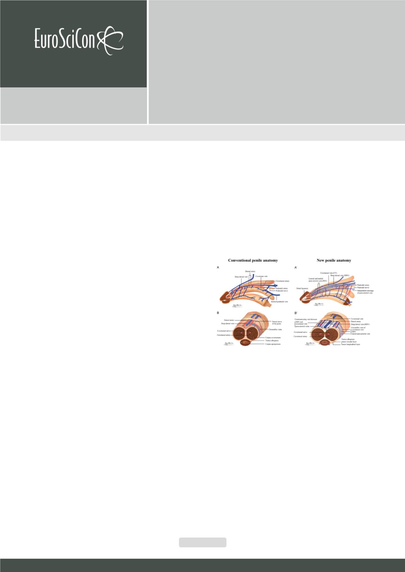
Figure 1:
Schematic illustration of conventional and new penile anatomy. (A)
Lateral view. The glans penis is exclusively composed of uniform sinusoids
only? The deep dorsal vein (DDV) is sandwiched in by a pair of dorsal arteries
(DA)? The 2:1 ratio of arteries to veins is the same as in the umbilicus vessel.
(B) Cross-section of a pendulous portion in the human penis. The tunica al-
buginea of the corpora cavernosa is consistently described as a one-layered
coat with uniform thickness. The median septum is complete. There is one
single DDV and two DAs between the tunica albuginea and Buck’s fascia.
Thus the penile vascular system still complies with the general anatomical
rule that veins number more than arteries do. In comparison, (A’) Lateral view:
The deep dorsal vein is consistently located in the median position and re-
ceives blood of the emissary veins from the corpora cavernosa and of the
circumflex vein from the corpus spongiosum. It is sandwiched between the
cavernosal veins, although these lie at a deeper position. Bilaterally, each dor-
sal artery is respectively sandwiched by its corresponding medial and lateral
para-arterial veins. Note that the lateral para-arterial vein merges with the me-
dial one proximally. The deeper color of the veins indicates the deepest part
of the vasculature. (B’) Cross section of the mid-penis. Note the number of
veins is seven, not one as was traditionally believed. (Although the number
becomes four at the level of the penile hilumbecause each pair of the nomen-
clature veins merges) Erection-related veins are arrayed in an imaginary arc
on the dorsal aspect of the tunica albuginea.
A physiological approach of penile venous stripping for
patients with erectile dysfunction on ambulatory basis
Geng Long Hsu
1, 2
1
Hsu’s Andrology, Taiwan
2
National Taiwan University, Taiwan
Geng Long Hsu, J Vasc Endovasc Therapy 2018, Volume 3
DOI: 10.21767/2573-4482-C1-002