


 43 / 65

Page 67
May 24-25, 2018
London, UK
Vascular Surgery 2018
3
rd
Edition of World Congress & Exhibition on
Vascular Surgery
Journal of Vascular and Endovascular Therapy
ISSN: 2573-4482
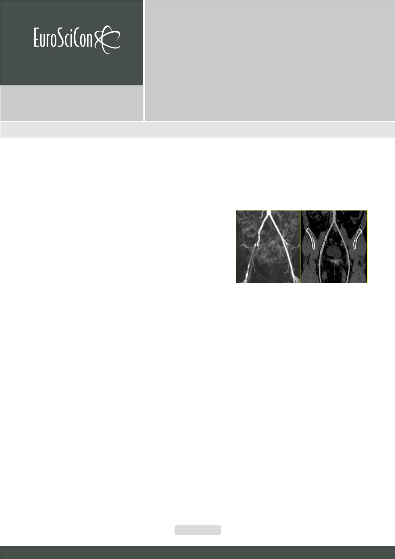
BACKGROUND:
PAOD is a clinical diagnosis, the clinical
Rutherford classification is based on walking impairment and the
extent of ischemic ulceration. However, the complete delineation
of the peripheral vascular tree ismandatory for treatment decision
and planning according to the TASC guidelines (1). For long time,
digital subtraction angiography was considered the gold standard
due to its high temporal and spatial resolution. However, DSA
is an invasive procedure requiring intra-arterial application of
contrast agents, with an associated risk of complications (2) and
the exposure of both patients and observers to ionizing radiation.
Thus, MR angiography and CT angiography have been developed
over the last 20 years in an effort to replace DSA for diagnostic
purposes and to limit it’s application to therapeutic procedures.
LEARNING OBJECTIVES:
This course will provide you with an
overview of MRA and CTA of the peripheral arteries with special
emphasis on their capabilities and limitations as well as most
recent technical developments (3-5). The following issues will be
covered:
• Volume coverage
• Spatial resolution
• Acquisition time
• Accurate and fast image post-processing
• Patient safety
• Cost efficiency
Recent Publications
1. Committee TS, Jaff MR, White CJ, Hiatt WR, Fowkes
GR, Dormandy J, et al. An Update on Methods for
Revascularization and Expansion of the TASC Lesion
Classification to Include Below-the-Knee Arteries: A
Supplement to the Inter-Society Consensus for the
Management of Peripheral Arterial Disease (TASC
II). J Endovasc Ther. 2015;22(5):663-77.Heilig M,
Egli M (2006) Pharmacological treatment of alcohol
dependence: Target symptoms and target mechanisms.
Pharmacology and therapeutics 111:855-876.
2. Waugh JR, Sacharias N. Arteriographic complications in
the DSA era. Radiology. 1992;182(1):243-6.
3. Edelman RR, Flanagan O, Grodzki D, Giri S, Gupta N,
Koktzoglou I. Projection MR imaging of peripheral
arterial calcifications. Magnetic Resonance in Medicine.
2015;73(5):1939-45.
4. Schreiner MM, Platzgummer H, Unterhumer S, Weber M,
Mistelbauer G, Groeller E, et al. Multipath Curved Planar
Reformations of Peripheral CT Angiography: Diagnostic
Accuracy and Time Efficiency. Cardiovascular and
interventional radiology. 2017.
5. Schreiner MM, Platzgummer H, Unterhumer S, Weber
M, Mistelbauer G, Loewe C, et al. A BMI-adjusted ultra-
low-dose CT angiography protocol for the peripheral
arteries-Image quality, diagnostic accuracy and radiation
exposure. Eur J Radiol. 2017;93:149-56.
Biography
Ruediger Schernthaner is an expert in cardiovascular imaging. He has been
developing CT angiography reformation techniques in collaboration with the
Technical University of Vienna and the Stanford Medical Center for more than
10 years. He has published more than 50 peer-reviewed publications in the
field of cardiovascular imaging and interventional oncology and authored the
chapter “Management of Peripheral Arterial Disease” in the book “Managing
Cardiovascular Complications in Diabetes”.
ruediger.schernthaner@meduniwien.ac.atState-of-the-art imaging of the peripheral vasculature
Ruediger E. Schernthaner
Medical University of Vienna, Austria
Ruediger E. Schernthaner, J Vasc Endovasc Therapy 2018, Volume 3
DOI: 10.21767/2573-4482-C1-002