


 14 / 16

Page 34
Insights in Enzyme Research
ISSN: 2573-4466
E u r o S c i C o n C o n g r e s s o n
Enzymology and
Molecular Biology
A u g u s t 1 3 - 1 4 , 2 0 1 8
P a r i s , F r a n c e
Enzymology 2018
Background:
Intervertebral disc (IVD) degeneration is a degenerative disease closely related to inflammation of nucleus pulposus
(NP) cells. Tumor necrosis factor alpha (TNF-α) is a pro-inflammatory cytokine which induces NP cells (NPCs) apoptosis and
accelerate IVD degeneration (IDD). The endoplasmic reticulum (ER) serves several important cell functions, which are essential
for normal cell function and survival. Nuclear factor-kappa B (NF-κB) is important for genes involved in cell survival, adhesion, and
proliferation. However, the roles of ER stress and NF-κB in IDD remain to be elucidated. This study aims to clarify the roles of NF-κB
and ER stress related unfolded protein response (UPR) in TNF-α-induced biological changes in rat NP cells and IVD degeneration.
Methods:
We cultured rat NPCs with different concentrations of TNF-α, with or without UPR and NF-κB pathway small interfering
RNA (siRNA). The protein levels of UPR markers (XBP1s and p-eIF2α) and p-p65 were measured by immunofluorescence staining
and Western blot analysis and were used to monitor UPR and NF-κB, respectively. Cell proliferation was evaluated by CCK-8 assay,
cell-cycle analysis and cyclin proteins expression. Apoptosis was detected by flowcytometry, TUNEL staining and Western blot
analyses. All the data were expressed as mean±SD and frommultiple independent experiments. The results were compared among
different groups by using unpaired student’s test. P-values<0.05 were considered significant.
Results:
TNF-α induced the apoptosis of some NPCs in the early stage and then accelerated the proliferation of surviving cells. In
addition, TNF-α stimulus up-regulated XBP1s, p-eIF2α and p-p65 at the protein level, which indicated that TNF-α activated UPR and
NF-κB signals in rat NP cells. However, these effects could be reversed by UPR and NF-κB siRNA, and UPR interference decreased
the expression of p-p65 notably. In parallel, both UPR and NF-κB interference reduced cell proliferation and enhanced apoptosis.
Conclusions:
Our study demonstrated that UPR reinforces the survival and proliferation of NPCs in TNF-α stimulus by activating
NF-κB signalling, which could be an important therapeutic target in the future.
Biography
Lu Chen has completed his Bachelor’s and Master’s degree in the School of Medicine, Southeast University and worked in Orthopaedic department as a Surgeon for 3 years. Now
he is further pursuing his PhD in Southeast University, and doing the basic research work in the field of Degenerative Disc Disease. He has publishedmore than 5 papers in reputed
journals. He has participated in the 12th International Congress of Chinese Orthopedic Association (COA), China E-Poster Presentation; International Symposium on Life Science
and Biological Engineering (ISLSBE), Hong Kong- Oral Presentation.
chan_dr8891@163.comUnfolded protein response exerts cytoprotection and
promotes the proliferation of nucleus pulposus cells in TNF-
α
stimulation by activating NF-
α
B
Lu Chen
1
, Lei Liu
1
, Zhi-Yang Xie
1
, Arjun Sinkemani
1
and Xiao-Tao Wu
1
Zhongda Hospital, Southeast University, Nanjing, China
Lu Chen et al., Insights Enzyme Res 2018, Volume 2
DOI: 10.21767/2573-4466-C1-002
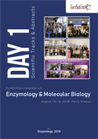
After treatment with different concentrations of TNF-α for
24h, the expression of UPR marker p-eIF2α was significantly
up regulated by Immunofluorescence staining
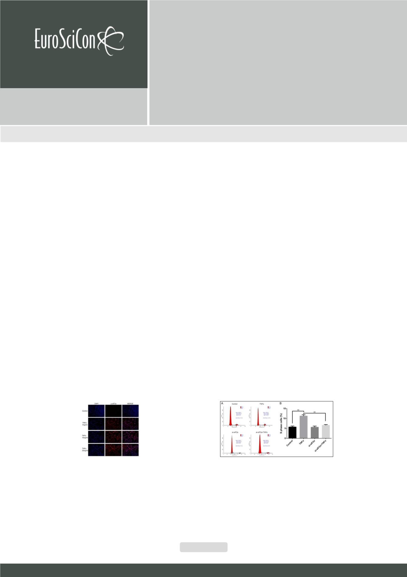
The effect of UPR on NPCs proliferation. (A) The percentage of S phase population cells
was measured by flowcytometry after eIF2α silencing under TNF-α stimulus. (B) eIF2α
interference reduced cell proliferation in TNF-α significantly. (*p < 0.05)