


 14 / 39

Volume 3, Issue 2
Insights in Analytical Electrochemistry
ISSN: 2470-9867
Analytical Chemistry-Formulation 2017
August 28-30, 2017
Page 28
8
th
Annual Congress on
&
14
th
International Conference and Exhibition on
August 28-30, 2017 Brussels, Belgium
Analytical and Bioanalytical Techniques
Pharmaceutical Formulations
Resonant X-ray emission spectroscopy for analysis of platinum anti-cancer complexes and their
interaction with biomolecules
Joanna Czapla-Masztafiak
1
, Jacinto Sá
2
, Jakub Szlachetko
3
and
Wojciech M Kwiatek
1
1
Institute of Nuclear Physics Polish Academy of Sciences, Poland
2
Uppsala University, Sweden
3
Institute of Physics, Jan Kochanowski University Kielce, Poland
Statement of the Problem:
The accidental discovery of the anticancer properties of cisplatin and its clinical introduction
in the 1970s paved the way for the use of platinum based metallodrugs in chemotherapy. Second-generation analogues (e.g.
carboplatin) were discovered shortly after. However, the clinical introduction of new anticancer metallodrugs has slowed down
dramatically, especially considering the number of new drugs synthesized each year. Probably, the most critical factor for this
slow progress is the inability to elucidate in a timely manner how metallodrugs induce tumor death, precluding the rational
development of new derivatives with enhanced anticancer capabilities. Most chemotherapeutic agents exert their antitumor
effect by damaging DNA and its replication machinery. Therefore, the correlation between the covalent bonding to DNA and
the cytotoxicity of the metal complex remains a central step in the search for new anticancer drugs.
Methodology & Theoretical Orientation:
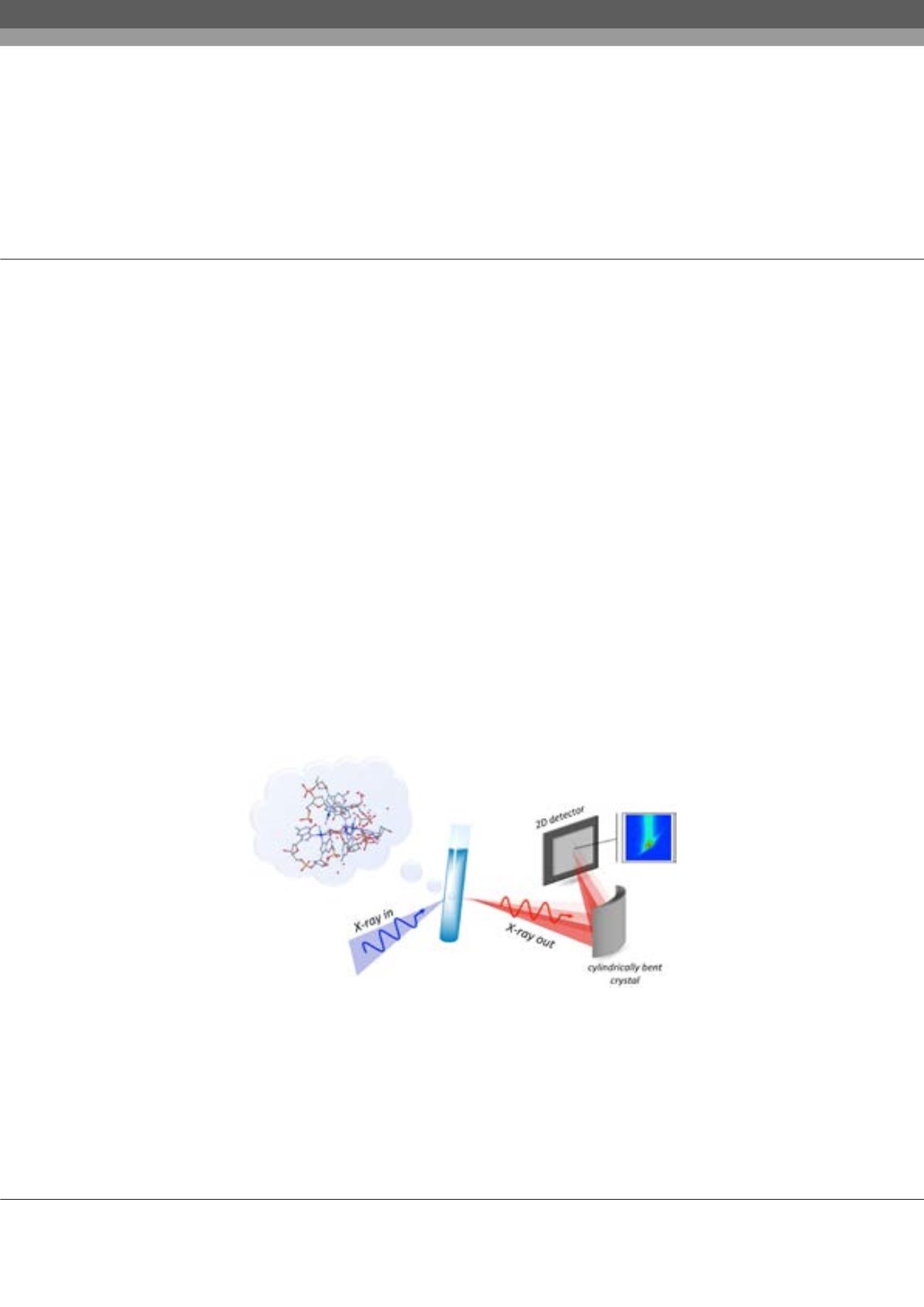
Herein, we report a strategy to follow the chemical structure and coordination of
platinum-based antitumor drugs by DNA under physiological conditions, namely by means of
in situ
resonant X-ray emission
spectroscopy (RXES). RXES is an atom specific photon-in photon-out scattering technique which avoids the tedious steps of
extraction and crystallization required by conventional X-ray techniques.
Conclusion & Significance:
The spectroscopic method proposed by us was successfully used to validate the mechanism of
action of cisplatin as well as elucidate the DNA binding of Pt103 compound that exhibits cytotoxic activity. Moreover, we
showed that RXES can be used to unveil the electronic structure of metallodrugs with high resolution and sensitivity and to
disentangle differences in the electronic structure of the metal canter induced by a secondary ligand stereochemistry.
Biography
Joanna Czapla-Masztafiak has her expertise in the use of X-ray spectroscopy to study biological systems. She obtained Master’s Degree in Medical Physics from
AGH University of Science and Technology in Krakow, Poland and PhD in Physical Sciences from Institute of Nuclear Physics Polish Academy of Sciences in
Krakow, Poland. She also successfully completed Postdoc project in Paul Scherrer Institute in Villigen, Switzerland. Her research interests cover the application
of X-ray spectroscopy to study DNA damage caused by radiation and chemical agents. Recently, she is developing laboratory instrument for X-ray absorption and
X-ray emission studies of the interaction of metal compounds with biomolecules.
joanna.czapla@ifj.edu.plJoanna Czapla-Masztafiak et al., Insights in Analytical Electrochemistry, 3:2
DOI: 10.21767/2470-9867-C1-002