


 4 / 23

Pharmacognosy 2019
March 11-12, 2019
London, UK
American Journal of Ethnomedicine
ISSN: 2348-9502
Page 40
Pharmacognosy and
Medicinal Plants
7
th
Edition of International Conference on
Nenad Stojiljković et al., Am J Ethnomed 2019, Volume 6
DOI: 10.21767/2348-9502-C1-009
Nanoliposome encapsulated lycopene ameliorates
methotrexate-induced hepatotoxicity
Nenad Stojiljković, Sonja Ilić, Nikola Stojanović, Milan Stoiljković
and
Slavica Stojnev
University of Nis, Serbia
Introduction:
Nano liposomes have the potential
to increase bioavailability, stability, improve time-
controlled drug releasing, enable cell specific
targeting and decrease adverse effects of drugs. In
this study, we evaluated the potential protective effect
of lycopene, a potent antioxidant carotenoid, given in
free and encapsulated form in methotrexate induced
hepatotoxicity in rats.
Methods:
Experiments were performed on 48 male
Wistar rats divided into eight groups of 6 animals,
treated daily by an intraperitoneal injection. MTX group
received methotrexate in a single dose (20 mg/kg) on
the first day; other experimental groups received the
same dose of methotrexate and empty nanoliposomes
(10 mL/kg) (MTX-NL-group), lycopene (6 mg/kg)
(MTX-LYC-group) and encapsulated lycopene (6 mg/
kg) (MTX-ENL-group), for 10 days. The remaining four
groups served as controls and received for 10 days:
corn oil (0.2 mL/day) (C-group), empty nanoliposomes
(10 mL/kg) (NL-group), lycopene (6 mg/kg) (LYC-group)
and encapsulated lycopene (6 mg/kg) (ENL-group).
Quantitative evaluation of structural and functional
changes of liver was performed by histopathological
(HE staining) and biochemical serum analyses and
determination of oxidative stress parameters.
Results:
Methotrexate induced severe functional and
morphological alterations of liver with conspicuous
disorganization of hepatic cords. Hepatocytes diffusely
exhibited apoptosis and degeneration with vacuolation
of the cytoplasm. Portal veins and sinusoid capillaries
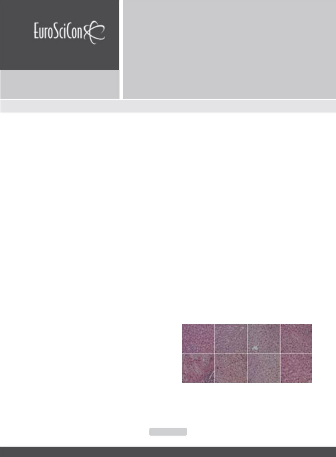
showed congestion. Marked inflammatory infiltratewas
present in the portal tract (Figure 1E). Pathohistological
findings were followed by AST and ALT increase and
disturbances of tissue antioxidant status. Application
of both forms of lycopene ameliorated changes in
serum AST and ALT and oxidative damage markers
and markedly reversed structural changes of liver
tissue induced by methotrexate. Animals that received
nanoliposome encapsulated lycopene showed higher
degree of recovery then those treated with free
lycopene in Figure 1.
Discussion:
Encapsulated lycopene was shown to
possess stronger antioxidant activity which could be
possibly related to its position in the lipid bilayer and its
higher stability in nanoliposomes which might prolong
the presence of lycopene in circulation. Treatment with
nanoliposome-encapsulated lycopene compared to free
lycopene has an advantage since it has more efficiently
reduced methotrexate induced hepatotoxicity.
Figure 1: Histological evaluation of liver tissue (HE, 400x) in: (A) C-group;
(B) NL-group; (C) LYC group; (D) ENL group; (E) MTX group; (F) MTX-NL
group; (G) MTX-LYC group and (H) MTX-ENL group