


 2 / 33

Page 27
conferenceseries
.com
Volume 3, Issue 2
ISSN: 2470-9905
Crystallography 2017
October 16-17, 2017
2
nd
International Conference on
October 16-17, 2017 | Chicago, USA
Applied Crystallography
Non-destructive studies of microstructure and elemental composition of crystalline materials through
energy-resolved neutron imaging
Anton S Tremsin
1
, Didier Perrodin
2
, Adrian S Losko
3
, Sven C Vogel
3
, Mark A M Bourke
3
, Gregory A Bizarri
2
, Edith D Bourret
2
, Takenao Shinohara
4
, Kenichi
Oikawa
4
, Winfried Kockelman
5
, S Ganguly6
and
Yan Gao
7
1
University of California at Berkeley, USA
2
Lawrence Berkeley National Laboratory, USA
3
Los Alamos National Laboratory, USA
4
Japan Atomic Energy Agency, Japan
5
STFC-Rutherford Appleton Laboratory, UK
6
Cranfield University, UK
7
GE Global Research, USA
E
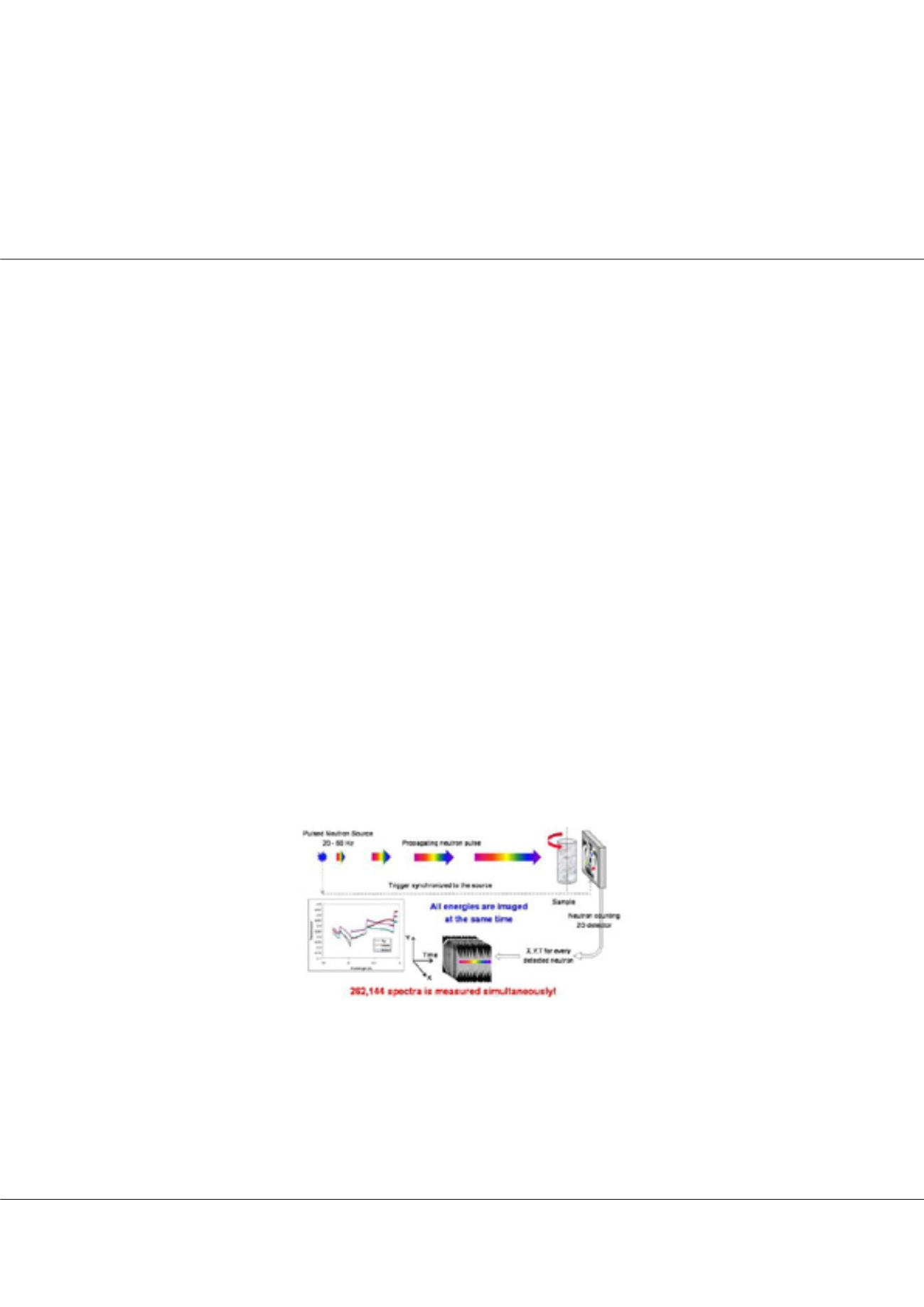
nergy-resolved neutron imaging provide unique possibilities to study materials non-destructively in situations, where other more
conventional techniques fail due to opacity of materials or their surrounding equipment (e.g., high temperature furnaces in case
of crystal growth). Microstructure of both polycrystalline and single crystal materials can be investigated due to the presence of Bragg
scattering of neutrons with wavelengths comparable to crystal lattice parameters. At the same time the elemental composition and
temperature of the material can be mapped remotely with ~0.1 mm resolution through the analysis of neutron resonance absorption
at epithermal energies, all from one measurement with no need to scan through the sample and thus allowing quantitative studies
of relatively slow dynamic processes, such as crystal growth. In this paper we demonstrate the unique capabilities of energy-resolved
neutron imaging to measure strain and some texture variation within metal welds, loaded fastener assemblies and metal samples
produced by additive manufacturing. In situ diagnostics of crystal growth parameters such as shape and location of liquid/solid
interface, mapping the elemental composition and visualization of macroscopic crystal defects and crystal mosaicity are also shown
for the growth of single crystal gamma scintillators. For some compound materials, such as Cs
2
LaLiBr
6
:Ce and BaBrCl:Eu, we directly
observed dynamics of phase separation within the liquid phase as well as dynamics of liquid/solid interface and dopant segregation
during crystal growth at 550
o
C and 850
o
C temperatures, respectively. These novel studies became possible with the recent progress
in novel high resolution neutron fast counting detectors and bright pulsed beamlines at spallation neutron sources, as well as
development of novel data analysis tools capable of processing hundreds of thousands neutron transmission spectra in acceptable
time, both of which will be briefly described in the paper.
Biography
Anton S Tremsin is currently working on the development of novel non-destructive testing techniques utilizing unique combination of high resolution event counting neutron
detectors and bright pulsed neutron sources. The detectors developed by him enable simultaneous detection of >250 thousand transmission spectra in each 55×55 µm2
pixel, enabling studies of microstructure of crystalline materials and mapping of their elemental composition, both ex situ and even in situ, as these materials are being grown
at high temperatures. A wide range of new state-of-the art experimental techniques in combination with data analysis tools have been demonstrated by him over recent
years in the field of materials science, structural engineering, single crystal growth and characterization, studies of magnetic phenomena, geosciences and many others,
result of which were presented at many international conferences and published in more than 200 research papers.
ast@ssl.berkeley.eduAnton S Tremsin et al., Struct Chem Crystallogr Commun, 3:2
DOI: 10.21767/2470-9905-C1-002
Figure-1: Schematic diagram of energy-resolved neutron imaging experimental setup