


 15 / 65

Page 35
May 24-25, 2018
London, UK
Vascular Surgery 2018
3
rd
Edition of World Congress & Exhibition on
Vascular Surgery
Journal of Vascular and Endovascular Therapy
ISSN: 2573-4482
C
onventional pharmaco-cavernosography including CT-
cavernosography provides little information on penile
venous anatomy, although it is acceptable in documenting veno-
occlusive erectile dysfunction (ED). We report an innovative
method, which can exclusively provide penile venous anatomy
for guiding penile venous stripping. From July 2010 to November
2017, 896 impotent men, aged 20 to 75 years, underwent this
methodof pharmaco-cavernosography inwhich twosetsof 60mL
of 50% omnipaque solution were administered intracavernously.
The first set of pilot cavernosograms was taken at intervals of
five, ten, twenty and thirty seconds after the commencement of
the injection. The second set of cavernosograms was taken
in the same intervals within 30 minutes following the pilot set,
preceded by the injection of 20 µg prostaglandin E1 (PGE1). For
comparison, the pilot cavernosograms were routinely performed
immediately postoperative on the patient undergoing penile
venous stripping. An analysis was conducted on the drainage
veins including deep dorsal vein (DDV), cavernosal veins
(CVs) and para-arterial veins (PAVs) accordingly. The veins
demonstrated in the pilot cavernosograms, and the second set
was compared in terms of venous numbers and presentation
percentage. A radio-opacity of the penile crura and that of the
femoral cortex was made. There was a statistically significant
difference (P<0.001) between the total number of independent
venous drainage channels and the presentation percentage of
DDV, CVs and PAVs observed in the pilot cavernosograms, and
those in second set (4.5 vs. 2.1; 97.48%, 60.35%, and 38.93%
vs. 57.08%, 29.37%, and 19.07%, respectively). A stronger radio-
opacity of the penile crura is unexceptional noted. Compared
with conventional pharmaco-cavernosography methods,
pilot cavernosograms are readily able to show detailed penile
venous anatomy which is indispensable for guiding venous
stripping surgery. It is, therefore, may be concluded that pilot
cavernosograms are an exclusively valuable addition
to
conventional of pharmaco-cavernosography and CT-c protocols
avernosography.
Recent Publications
1. Hsu G L, Chen H S, Hsieh C H, Lee W Y, Chen K L and
Chang C H (2010) Clinical experience of a refined
penile venous stripping surgery procedure for patients
with erectile dysfunction: is it a viable option? Journal
of Andrology 31:271-280.
2. Hsu G L, Chen H S, Hsieh C H, Lee W Y, Chen K L and
Chang C H (2010) Salvaging penile venous stripping
surgery. Journal of Andrology 31:250-260.
3. Hsieh CH, Liu S P, Hsu G L, Chen HS, Molodysky E, Chen
Y H and Yu H J (2012) Advances in our understanding
of mammalian penile evolution, human penile anatomy
Novel pilot films providing indispensable information in
pharmaco-cavernosography
Chi Can Huynh
1
and
Geng Long Hsu
2, 3
1
The Male Clinic, Australia
2
Hsu’s Andrology, Taiwan
3
National Taiwan University, Taiwan
Chi Can Huynh et al., J Vasc Endovasc Therapy 2018, Volume 3
DOI: 10.21767/2573-4482-C1-002
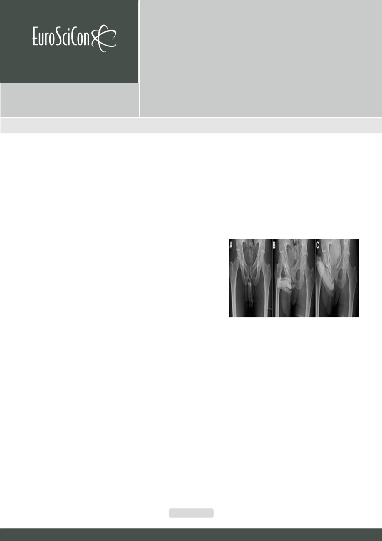
Figure 1:
Dual pharmaco-cavernosography. (A) An anterior-posterior
view was first used for the first set of our dual cavernosography. The
complex distribution of the erection related veins was characteristic.
Note at least 7 significant veins drained sinusoidal blood away from
the corpora cavernosa. (B) A 30-degree right oblique position was
then used to see the lateral view of the erection related veins. (C) A
pharmacocavernosogram showed a veno-occlusive dysfunction
(VOD) despite the rigid erection exist 15 minutes after artificially in-
duced erection by intracavernous prostaglandin E1