


 6 / 25

Vascular Surgery 2019
Journal of Vascular and Endovascular Therapy
ISSN: 2573-4482
Page 36
March 28-29, 2019
Rome, Italy
Vascular Surgery
4
th
Edition of World Congress & Exhibition on
Can carotid endarterectomy only be indicated by Doppler
ultrasonography?
Fatima El Hajj
and
Christiano Stchelkunoff Pecego
IAMSPE, Brazil
Fatima El Hajj et al., J Vasc Endovasc Therapy 2019, Volume 4
DOI: 10.21767/2573-4482-C1-005
S
troke is a disease of great health relevance due to its
morbidity and high costs generated. Symptomatology
varies from asymptomatic, fugitive amaurosis, transient
ischemic attack (TIA) and direct manifestations of stroke.
In view of this, prevention would be the best option to
reduce costs secondary to morbidity and mortality due
to stroke. Carotid endarterectomy (ACE) is the most
common preventive procedure. ECA is a surgical technique
consolidated 50 years ago. Large studies such as NASCET,
VA, ECST, ACAS and ASCT have already analyzed and
proven the indications, cost-effectiveness and limitations
of the technique. Digital angiography is the gold standard
for determining the degree of carotid stenosis, as methods
of investigating carotid stenosis and defining the surgical
indication of endarterectomy. Because it is an invasive
examination, with a risk ofmajor complications (TIA/stroke)
of approximately 4%, it has been progressively replaced by
tomography (Angio-CT) or resonance angiography (Angio-
NMR). At the same time, Doppler ultrasonography (US) is a
non-invasive,low-costmethodofscreeningcarotidstenosis.
With the incessant progress of diagnostic methods,
Doppler ultrasonography (US) has proven to be a method
of choice for noninvasive evaluation of the carotid arteries.
The degree of carotid artery stenosis is largely based on
either a peak systolic velocity or final diastolic velocity
analysis, or both, of the carotid artery. Doppler scanning in
pulsed mode combined with B-mode ultrasound allows the
diagnosis of carotid atheromatous lesions (>70% stenosis),
with sensitivity and specificity above 90%. The US adds
comparative advantages to the other contrasted methods,
since it is a lower cost procedure, it lacks complications and
contraindications, it is easy to access, it does not require
the use of contrast, and it also has information about plaque
morphology, stenosis percentage and topography of the
carotid bifurcation.
Recent Publications
1. Brunno Cezar Framil Sanchez, Lineu Amaro
Rodrigues Junior, Felipe Trentin Neves,
Thiago Correa Tambelli, Fernando Eduardo
Paulatti Frederico, Fatima Mohamad El Hajj,
Juliana Monteiro de Abreu, Tatiana Milunovic
Lobo Rosa, Alexis Iury Framil Sanches and
Antônio Alberto Ramos Argento (2011) Giant
condyloma. Journal of the Faculty of Medical
Sciences of Sorocaba 13:25-27.
Biography
Fatima El Hajj is a Brazilian – Lebanese Vascular Surgeon. MD in Pontif-
ical Catholic University of São Paulo – PUCSP. General Surgery degree
inMunicipal Public Server Hospital of São Paulo – HSPM. Vascular Sur-
gery Degree in Estadual Public Server of São Paulo – IAMSPE. Vascular
Surgeon at AnaliaMed Health and Wellness. CEO AnaliaMed Diagnose
Center.
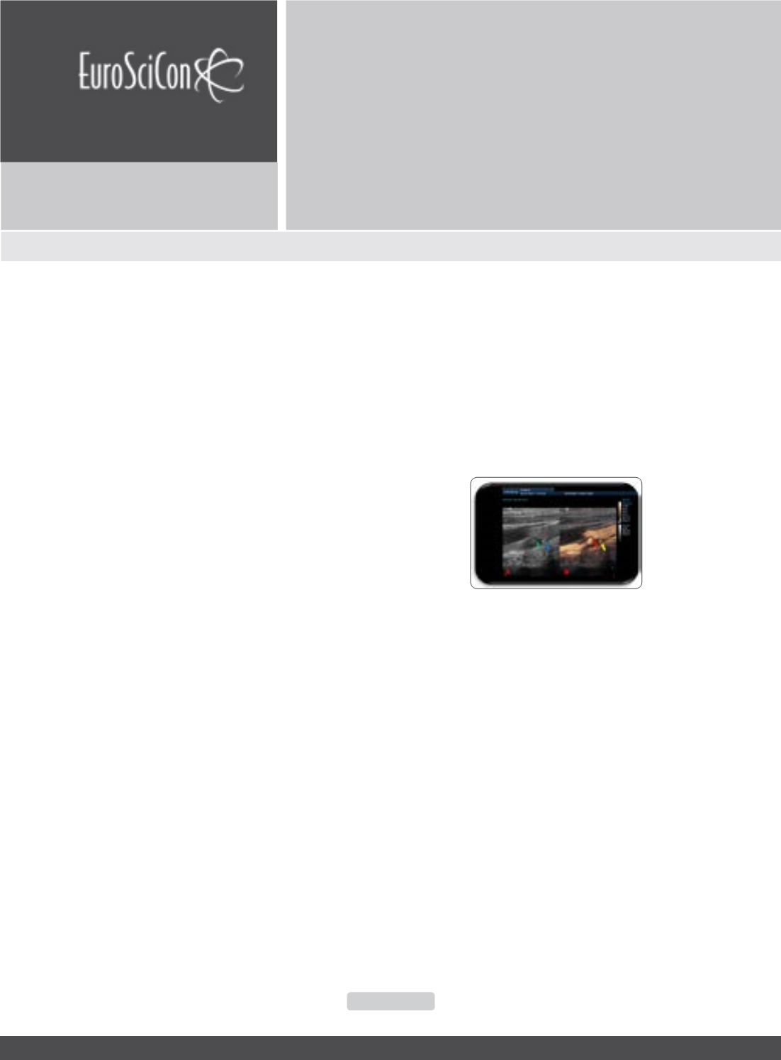
fatimamedlv@hotmail.comFigure 1:
Diagnostic complementation with CEUS technique..