ISSN : 2349-3917
American Journal of Computer Science and Information Technology
Input Of Raman, SERS And Fluorescence Spectroscopy To Nanomedicine Research
EuroSciCon Joint Event on Laser Optics & Photonics and Atomic & Plasma Science
July 16-17, 2018 Prague , Czech republic
Igor Chourpa, Lynda Miloudi, Franck Bonnier, Katel Herve-Aubert, Mathias Pacaud, Emilie Allard-Vannier, Emilie Munnier, Anastasia Ignatova, Amir Fahmi and Alexey Feofanov
University of Tours, Tours, France Rhein-Waal University of Applied Sciences, Germany Shemyakin-Ovchinnikov IBCh RAS, Russia Lomonosov Moscow State University, Russia
ScientificTracks Abstracts: Am J Compt Sci Inform Technol
DOI: 10.21767/2349-3917-C1-002
Abstract
Research in nanomedicine is known to be a stimulating field for fruitful interaction between complementary scientific expertises. Indeed, it integrates the most recent advances in material chemistry, medicinal chemistry, physiology, biotechnology and biophysics. In the present talk, we will describe how the nanomedicine research can take benefit from the advanced molecular optical spectroscopy and spectral imaging. Conventional Raman and Surface-Enhanced Raman Scattering (SERS) are analytical techniques attracting a close attention due to several advantages they offer, namely direct analysis of many complex samples, with high spatial resolution and high molecular specificity, at relatively low cost. In case of SERS and fluorescence, increased selectivity and sensitivity are additional advantages. Furthermore, as we demonstrate, it is possible to combine SERS with fluorescence, either in a frame of a complementary analysis or via the simultaneous co-detection of both scattered and emitted photons. Such a combination/coupling allows, for instance, a more relevant interpretation of the molecular state and environment of active ingredients or of nanomedicine platforms in cells and tissues. Nevertheless, spectroscopic analysis of biological samples leads to collection of complex datasets often subjected to strong variations. It is particularly the case for spectral imaging, with thousands of spectra recorded when mapping the region of interest. For a more efficient mining of spectroscopic data, chemometric approaches are necessary. The input of our advanced spectroscopic approaches to nanomedicine research will be illustrated using their applications to bioanalytics (study of active molecules in pharmaceutic/cosmetic nanoforms and in biological cells and tissues) as well to diagnostics (development of novel nanoprobes for multimodal biomedical imaging of cancers). As we show, this input is not limited to characterize/evaluate nanomedicine forms, but it is extended to create novel spectroscopy-based diagnostic approaches, expected to be more specific, sensitive and reliable. Recent Publications 1. Gautier J, Allard-Vannier E, Munnier E, Souce M and Chourpa I (2013) Recent advances in theranostic nanocarriers of doxorubicin based on iron oxide and gold nanoparticles. J Control Release 10;169 (1-2):48-61. 2. Gautier J, Allard-Vannier E, Burlaud-Gaillard J, Domenech J and Chourpa I (2015) Efficacy and Hemotoxicity of Stealth Doxorubicin-Loaded Magnetic Nanovectors on Breast Cancer Xenografts. J Biomed Nanotechnol 11(1):177-89.
Biography
Igor Chourpa has completed his PhD from the University of Reims, France in 1996. He joined the University of Tours in 1997. He is the Director of Nanomedicine and Nanoprobes laboratory (EA6295). This interdisciplinary team develops bio-analytical methods and nanomedicine technology for drug delivery and disease diagnostics. His specialization is in Molecular Optical Spectroscopy (Raman, SERS, fluorescence and IR) and Spectral Imaging. He has published more than 60 papers in reputed journals and 2 book chapters. He has experience in coordination of national and international research projects and has been acted as expert for numerous calls. He is an Editorial Board Member of Journal of Analytical Methods in Chemistry.
E-mail: igor.chourpa@univ-tours.fr
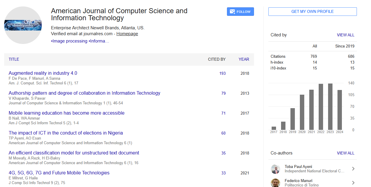
Google Scholar citation report
Citations : 769
Abstracted/Indexed in
- Google Scholar
- Genamics JournalSeek
- China National Knowledge Infrastructure (CNKI)
- CiteFactor
- Open Academic Journals Index (OAJI)
- Directory of Research Journal Indexing (DRJI)
- Jour Informatics
- CiteSeerx
- Journal Index.net
- Secret Search Engine Labs
Open Access Journals
- Aquaculture & Veterinary Science
- Chemistry & Chemical Sciences
- Clinical Sciences
- Engineering
- General Science
- Genetics & Molecular Biology
- Health Care & Nursing
- Immunology & Microbiology
- Materials Science
- Mathematics & Physics
- Medical Sciences
- Neurology & Psychiatry
- Oncology & Cancer Science
- Pharmaceutical Sciences