ISSN : 2576-392X
Dentistry and Craniofacial Research
Temporomandibular Joint Ankylosis Pattern, Causes and Management among a Sample of Sudanese Children
Yousif I Eltohami*, Amal H Abuaffan, Riham A Alsagh, Ethar A Abdalgadir, Hisham H Mohamed and Asaad K Ali
University of Khartoum, Faculty of Dentistry, Sudan
- *Corresponding Author:
- Amal H Abuaffan
University of Khartoum, Faculty of dentistry, Sudan
Tel: +249912696035
E-mail: amalabuaffan@yahoo.com
Received date: January 25, 2017; Accepted date: May 05, 2017; Published date: May 12, 2017
Citation: Eltohami YI, Abuaffan AH, Alsagh RA, et al. Temporomandibular Joint Ankylosis Pattern, Causes and Management among a Sample of Sudanese Children. Den Craniofac Res.Vol.2 No.1:6. doi: 10.21767/2576-392X.100006
Abstract
Background: Temporomandibular joint ankylosis is the fusion of mandibular condyle to the glenoid fossa, which causes distressing conditions. It may be due to trauma or infection. The aim of this study is to determine the frequency of TMJ Ankylosis in Khartoum teaching dental hospital among children.
Materials and methods: A retrospective cross sectional study for 48 patients (32 male, 16 female) aged 0-18 years old. Data were collected from patients records registered from January 2009 to April 2015.
Results: Males were more affected than females, the most affected age group was 7-12 years old, bilateral ankylosis more common than unilateral, intra capsular ankylosis is the most common type, and micrognathia is the most common deformity. 41 patients received treatment, coronoidectomy with condylectomy and physiotherapy is the most preferable treatment method. Also Condylectomy + Gap arthroplasty + Physiotherapy and Gap arthroplasty + Coronoidectomy + Physiotherapy were used. In some cases physiotherapy overcome the ankylosis, and only 7 patients didn’t receive any type of treatment.
Conclusion: The prevalence of TMJ ankylosis among children was high; the most common causes were trauma and infection, whereas most of patients came with intracapsular type ankylosis in children leads to facial deformities. Improvement of awareness regarding condyle fracture is required.
Keywords
Temporomandibular joint; Ankylosis; Condyle fracture
Introduction
Temporomandibular joint (TMJ) is a synovial diarthrodial joint formed between the condyle of the mandible and the articular tubercle of temporal bone and articular fossa [1].
TMJ Ankylosis is fusion of the mandibular condyle to the glenoid fossa in the base of the skull, which causes distressing conditions such as impaired speech, difficulty in chewing, poor oral hygiene, facial disfigurement, compromise of the airway, and psychological stress [2].
Various etiological factors had been attributed to TMJ ankylosis; trauma, local and systemic inflammatory conditions, neoplasm, and TMJ infection. The most frequent one trauma and infection [3].
Fractures of the condylar head are more prone to postoperative ankylosis of the TMJ, and that the possible risk factors seem to include the technique used for fixation and damage to the disc, together with an anterior mandibular fracture and with the remaining fractured fragment [4].
Bilateral TMJ ankylosis caused by systemic infection is reported [4]. The clinical findings of TMJ ankylosis in children in unilateral ankylosis reveal unilateral hypoplasia of the mandible and deviation of the chin to the affected side. Bilateral ankylosis results in bird-face appearance; night snoring and obstructive sleep apneas are the other clinical findings in bilateral ankylosis [5]. Later joint involvement after 15 years of facial deformity is marginal or nil but functional loss is severe [6].
TMJ imaging comprise; plain radiography, panoramic radiography, tomograms, conventional CT, arthrography, three dimensional CT, magnetic resonance imaging, ultrasonography, and radionuclide imaging [7,8].
The management goal in TMJ ankylosis is removal of the ankylotic mass, restoring the form and function of the joint, mouth opening, relief of upper airway obstruction, and prevention of recurrence [9].
A number of surgical approaches have been devised to restore normal joint functioning and prevent re-ankylosis. Three basic techniques are used; gap arthroplast, interposition arthroplasty and joint reconstruction [10,11].
Methodology
Ethical approval was obtained from University of Khartoum Faculty of Dentistry, Ethical committee review board, and from research unit at Khartoum Teaching Dental Hospital to conduct this study.
This retrospective cross-sectional hospital based study was carried out to determine the pattern of TMJ ankylosis in KTDH among children, it consist of patients who were diagnosed with TMJ ankylosis and underwent surgical treatment in the period January 2009 to April 2015, this study conducted for Sudanese patients under 18 years old and diagnosed with TMJ ankylosis, and patients who had TMJ disorder other than ankylosis or their records were incomplete were excluded.
Total coverage of all patients records from January 2009 to April 2015 who fulfilled inclusion and exclusion criteria were included accordingly the sample size was 48 patients.
The following information had been obtained from the patient records, gender, age, type of ankylosis, side of ankylosis, mouth opening during theatre, growth deformity and treatment modality.
Data was collected, summarized, coded and entered to the Statistical Package for Social Sciences (SPSS) program (version 20) in the computer. Frequency distribution tables, graph were used to represent the results.
Results
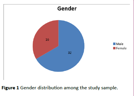
A total of 48 patients were included in this study, according to their gender result showed that 66.6% (n=32) were males while 33.4% (n=16) were female Figure 1.
The age of the patients ranged from 0 to 18 years old the highest percentage group was from 7-12 years 44% shown in Table 1.
| Age | Number | Percentage |
|---|---|---|
| 0 to 6 years | 9 | 19% |
| 7 to 12 years | 21 | 44% |
| 13 to 18 years | 18 | 37% |
| Total | 48 | 100% |
Table 1: Shows the age distribution among the study sample.
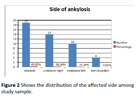
The study revealed that bilateral ankylosis was the predominant one account (40%), and the unilateral ankylosis on the right side (30%) was more predominant than the left side (21%) shown in Figure 2.
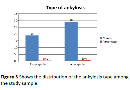
According to the types the majority of patients presented with intracapsular type 60% shown in Figure 3.
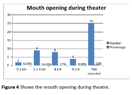
The intra-operative mouth opening achieved ranged from 0 cm to 8 cm, the predominant reading was from 2.1 cm to 4 cm were 19% as seen in Figure 4.
According to the study micrognathia was the predominant growth deformity 19% as shown in Table 2. Treatment was done to 41 patients, the most common treatment modality used was Coronoidectomy + Condylectomy + Physiotherapy for 21%, 15% of patients had Gap arthroplasty + Physiotherapy and 15% didn’t receive any treatment refer Table 3.
| Growth deformity | Number | Percentage |
|---|---|---|
| Retrognathia | 7 | 15% |
| Micrognathia | 9 | 19% |
| Retrognathia + Micrognathia | 3 | 6% |
| Not recorded | 29 | 60% |
Table 2 Shows the growth deformities among study sample.
| Treatment | Number | Percentage |
|---|---|---|
| Gap arthroplasty + Physiotherapy | 7 | 15% |
| Interpositional gap arthroasty + Physiotherapy | 0 | 0% |
| Coronoidectomy + Physiotherapy | 5 | 10.00% |
| Condylectomy + Physiotherapy | 6 | 12.50% |
| Coronoidectomy + Condylectomy + Physiotherapy | 10 | 21% |
| Physiotherapy | 3 | 6.00% |
| Condylectomy + Gap arthroplasty + Physiotherapy | 6 | 12.50% |
| Gap arthroplasty + Coronoidectomy + Condylectomy + Physiotherapy | 1 | 2% |
| Interpositional gap arthroplasty + Coronoidectomy + Condylectomy + Physiotherapy | 1 | 2% |
| Gap arthroplasty + Coronoidectomy + Physiotherapy | 2 | 4% |
| No treatment done | 7 | 15.00% |
Table 3 Shows the types of treatment recorded among study sample.
Discussion
The aim of this study was to determine the frequency of TMJ ankylosis in Khartoum Teaching Dental Hospital among children according to; age, type, cause, severity, treatment and the side involved.
Based on the study the mean age of the sample was 11.5 years which coincides with the studies by Madjidi and Bala [12,13]. The result was in contrast with He, Ellis and Zhang findings in which the mean age was 23 years [12-14].
Male to female ratio in the study was 2:1 Based on the study most patients presented with unilateral ankylosis with bilateral ankylosis presenting to lesser degree which coincides with He, et a. results and Bala results [13,14].
The cause of ankylosis in the majority of patients in the study was duo to trauma which coincides with Bala findings, and contrast with Madjidi A. findings in which the majority of the cases were duo to infection [12,13].
In this study, the mean mouth opening achieved during theater was 4.1 cm which coincides with Bob Rishiraj et al. findings also in contrast with UM Das et al. results and Rahul Hegde et al. findings in which a mouth opening of 2.5 cm was achieved [15-17].
The patients in the study dominantly presented with micrognathic deformities which coincide with de Castro e Silva et al. results, but it is in contrast with Liaqat et al. results in which the patient presented with retrognathia and Rahul Hegde et al. findings in which the patient presented with both micrognathia and retrognathia [17-19].
In this study the most used treatment was Coronoidectomy + Condylectomy + Physiotherapy, also Condylectomy + Gap arthroplasty + Physiotherapy which coincide with Orhan Gu¨ven findings [20].
Conclusion
TMJ ankylosis in children is a challenging problem affecting the mandible resulting in esthetic and functional deformities. The most common causes are trauma and infection. Males are affected more than females. Ankylosis in children leads to facial deformities such as micrognathia which is more common than retrognathia. Majority of patients presented with unilateral ankylosis. intracapsular TMJ ankylosis is found to be more prevalent than extracapsular. Coronoidectomy + condylectomy + physiotherapy is the most used protocol for treatment, which lead to improvement in mouth opening with a mean of 4.1 cm during surgery which can be increased by using aggressive physiotherapy.
Recommendation
The researchers recommended improvement of the awareness regarding condylar fracture, early intervention by distraction osteogenesis to reduce the psychological impact of jaw deformities, continuous follow up to the patients after surgery to prevent recurrence and the surgeons in Sudan should learn about new surgical skills and technique to reduce the post-operative complications.
References
- Bath-Balogh M, Fehrenbach M (2011) Illustrated dental embryology, histology and anatomy (4thedn), Elsevier Saunders, St. Louis, Missouri, USA.
- Anyanechi CE, Osunde OD, Bassey GO (2015) Use of oral mucoperiosteal and pterygo-masseteric muscle flaps as interposition material in surgery of temporomandibular joint ankylosis: A Comparative Study. Ann Med Health Sci Res 5: 30-35.
- Vasconcelos BC, Porto GG, Bessa-Nogueira RV (2008) Temporo mandibular joint ankylosis. Rev Bras Otorrinolaringol 74: 34-38.
- Xiang G, Long X, Deng M, Han Q, Meng Q, et al. (2014) A retrospective study of temporomandibular joint ankylosis secondary to surgical treatment of mandibular condylar fractures. Br J Oral Maxillofac Surg 52: 270-274.
- Zarb GA (1995) Temporomandibular joint and masticatory muscle disorders (2ndedn), Elsevier Inc, St. Louis, Missouri, USA.
- deLeeuw R, Klasser GD (2013) American academy of orofacial pain: Guidelines for assessment, diagnosis, and management eds. (5thedn), Quintessence Publishing, Chicago, USA
- Sayan NB, Karasu HA, Uyank LO, Aytaç D (2007) Two-stage treatment of TMJ Ankylosis by early surgical approach and distraction Osteogenesis. J Craniofac Surg 18: 212-217.
- Felstead AM, Revington PJ (2011) Surgical management of temporomandibular joint ankylosis in ankylosing spondylitis. Int J Rheumatol 2011: 1-5.
- Dimitroulis G (2004) The interpositional dermis-fat graft in the management of temporomandibular joint ankylosis. Int J Oral Maxillofac Surg 33: 755-760.
- Lei Z (2002) Auricular cartilage graft interposition after temporomandibular joint ankylosis surgery in children. J Oral Maxillofac Surg 60: 985-987.
- Majumdar A, Bainton R (2004) Suture of temporalis fascia and muscle flaps in temporomandibular joint surgery. Br J Oral Maxillofac Surg 42: 357-359.
- Madjidi A, Briat B, Couly G (1994) Temporomandibular ankylosis in children: Apropos of 30 cases. Rev Stomatol Chir Maxillofac 95: 157-160.
- Balaji S (2010)Bifid mandibular condyle with temporomandibular joint ankylosis: A pooled data analysis. Dent Traumatol 26: 332-337.
- He D, Ellis E, Zhang Y (2008) Etiology of Temporomandibular joint Ankylosis secondary to Condylar fractures: The role of concomitant Mandibular fractures. J Oral Maxillofac Surg 66: 77-84.
- Rishiraj B, Mc-Fadden L (2001) Treatment of temporomandibular joint ankylosis: A case report. J Can Dent Assoc 67: 659-663
- Das U, Keerthi R, Ashwin D, VenkataSubramanian R, Reddy D, et al. (2009) Ankylosis of temporomandibular joint in children. J Indian Soc Pedod Prev Dent 27: 116-120.
- Hegde R, Devrukhkar V, Khare S, Saraf T (2015) Temporomandibular joint ankylosis in child: A case report. J Indian Soc Pedod Prev Dent 33: 166-169.
- de Castro e Silva LM, Pereira FVA, Vieira EH, Gabrielli MF (2011) Tracheostomy-dependent child with temporomandibular ankylosis and severe micrognathia treated by piezosurgery and distraction osteogenesis: Case report. Br J Oral Maxillofac Surg 49: e47-9.
- Liaqat S, Baig AM, Bukhari SG, Ahmed W (2011) Distraction osteogenesis of a unilateral hypoplastic mandible. J Ayub Med Coll Abbottabad 23: 174-176.
- Güven O (2004) Treatment of temporomandibular joint ankylosis by a modified fossa prosthesis. J Craniomaxillofac Surg 32: 236-242.
Open Access Journals
- Aquaculture & Veterinary Science
- Chemistry & Chemical Sciences
- Clinical Sciences
- Engineering
- General Science
- Genetics & Molecular Biology
- Health Care & Nursing
- Immunology & Microbiology
- Materials Science
- Mathematics & Physics
- Medical Sciences
- Neurology & Psychiatry
- Oncology & Cancer Science
- Pharmaceutical Sciences
