ISSN : ISSN: 2576-1455
Journal of Heart and Cardiovascular Research
Superior Vena Cava Syndrome
1University of Pittsburgh Medical Center, Pittsburgh, Pennsylvania, USA
2Harlem Hospital Center, New York, New York, USA
- *Corresponding Author:
- Deepak Kumar Pasupula
University of Pittsburgh Medical Center
9100 Babcock Blvd, Pittsburgh
Pennsylvania 15237, USA
Tel: 412-999-8606
E-mail: 84pdeepak@upmc.edu
Received Date: June 11, 2018; Accepted Date: June 22, 2018; Published Date: June 29, 2018
Citation: Pasupula DK, Malleshappa SKS, Terla V (2018) Superior Vena Cava Syndrome. J Heart Cardiovasc Res Vol.2 No.1:5
Copyright: © 2018 Pasupula DK, et al. This is an open-access article distributed under the terms of the creative commons attribution license, which permits unrestricted use, distribution and reproduction in any medium, provided the original author and source are credited.
Keywords
Superior vena cava syndrome; Schamroth’s sign; Small-cell lung carcinoma
Image Article
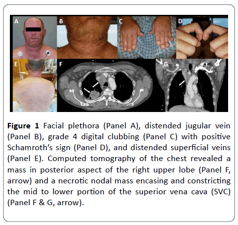
A 49-year-old male with 68 pack-year history of smoking tobacco and 10 cannabis cigarettes per month for 29 years, presented with bilateral upper extremity swelling and rightsided chest pain (Figure 1). On examination, he had facial plethora (Panel A), distended jugular vein (Panel B), grade 4 digital clubbing (Panel C) with positive Schamroth’s sign (Panel D), and distended superficial veins (Panel E). Computed tomography of the chest revealed a mass in posterior aspect of the right upper lobe (Panel F, arrow), and a necrotic nodal mass encasing and constricting the mid to lower portion of the superior vena cava (SVC) (Panel F & G, arrow). Diagnosed with small-cell lung carcinoma (SCLC) on biopsy. SCLC is second most common malignant cause for SVC syndrome. Severity of signs and symptoms reflects the degree and rate of SVC narrowing. Digital Clubbing is a common finding in lung cancer, but rarely due to SCLC. He was started on carboplatin and etoposide, after 2nd cycle he developed pancytopenia and acute respiratory distress syndrome. Considering overall clinical complexity, family opted for palliative management. He died in 6 weeks from the diagnosis.
Figure 1: Facial plethora (Panel A), distended jugular vein (Panel B), grade 4 digital clubbing (Panel C) with positive Schamroth’s sign (Panel D), and distended superficial veins (Panel E). Computed tomography of the chest revealed a mass in posterior aspect of the right upper lobe (Panel F, arrow) and a necrotic nodal mass encasing and constricting the mid to lower portion of the superior vena cava (SVC) (Panel F & G, arrow).
Open Access Journals
- Aquaculture & Veterinary Science
- Chemistry & Chemical Sciences
- Clinical Sciences
- Engineering
- General Science
- Genetics & Molecular Biology
- Health Care & Nursing
- Immunology & Microbiology
- Materials Science
- Mathematics & Physics
- Medical Sciences
- Neurology & Psychiatry
- Oncology & Cancer Science
- Pharmaceutical Sciences

