Studying the Effect of Gamma Irradiation on Bovine Offal's Infected with Mycobacterium tuberculosis Bovine Type
1Department of Food Control, Benha University, Qalubiya Governorate Egypt
2Animal Health Research Institute, Giza Governorate, Egypt
3Radioisotope Department Nuclear Research Center, Atomic Energy Authority, Egypt
4General Organization for Veterinary Services, Giza Governorate, Egypt
- *Corresponding Author:
- Shaltout FA
Faculty of Veterinary Medicine
Department of Food Control
Benha University, Benha
Qalubiya Governorate, Egypt
Tel: 0020132461411
E-mail: fahimshaltout@hotmail.com
Received Date: October 23, 2017; Accepted Date: November 01, 2017; Published Date: November 09, 2017
Citation: Shaltout FA, Riad EM, TES Ahmed, Asmaa AA (2017) Studying the Effect of Gamma Irradiation on Bovine Offal's Infected with Mycobacterium tuberculosis Bovine Type. J Food Biotechnol Res. Vol.1 No.1:6
Abstract
The presence of microbial pathogens on human foods is a serious global Problem even in highly industrialized and developed countries. The awareness of foodborne diseases by consumers will increase, and therefore, there is a pressure to improve the safety of the food supply. Gamma ray is highly effective in inactivating microorganisms in various foods and offers a safe alternative method of food decontamination. In the present study, a total of 35 samples from T.B. infected carcasses 15 samples of offal's ((7) liver and (8) Kidney) and 15 samples from different lymph nodes ((10) Hepatic and (5) Renal)) were collected from some governmental Egyptian abattoirs confirmed to be infected with Mycobacterium tuberculosis bovine type by Real Time PCR were experimental treated with 0.0, 2.5, 5 and 7.5 kGy of gamma rays then, reexamined using RT-PCR for Mycobacterium tuberculosis bovine type infection. The results indicated that the reduction rate is decreased by increase the dose level of Gamma rays. At 0.0 kGy all samples still 100% infected and 46.6% still infected at 2.5 kGy and 20% still infected at 5 kGy and, At 7.5% all examined samples are failed to be detected of T.B. Infected offal's. Moreover, the examined samples of T.B. Infected lymph nodes showed that at 0 kGy all samples still 100% infected and 53.3% still infected at 2.5 kGy and 13.3% still infected at 5 kGy and, at 7.5% all examined samples are free from mycobacterium infection. The effect on (Color and Odor and Texture) parameter after exposure to Gamma rays on T.B. infected samples proved that most of tested samples have slight changes in color (pale color), odor (characteristic odor of irradiation) and texture (friable) in the first 24 hours and all tested samples have been returned back into the normal parameter after 1 week. The results of the present study showed that it's advisable to use the Gamma irradiation for saving a huge amount of condemned meat due to T.B infected cattle carcasses and using it as low grade meat.
Keywords
Mycobacterium bovis; Bovine offal's; Gamma irradiation
Introduction
The presence of microbial pathogens in human foods is a serious global problem. Even in highly industrialized and developed countries like the United States, pathogen-contaminated foods and the resulting health and economic impacts are significant. According to CDC [1], each year Americans suffer 76 million infections, 325,000 hospitalizations, and approximately 5,000 deaths due to pathogen-contaminated foods. These events carry an estimated annual healthcare cost totaling 7 billion $ [2]. Consider also that more than 74 million lb of pathogencontaminated meat and meat products were recalled between 2000 and 2003 [3], and the need for pathogen reduction is clear.
Safety and efficiency of food irradiation have been approved by several authorities (FDA, USDA, WHO, FAO, etc.) and scientific societies based on extensive research [4,5]. However, market success of irradiated foods has not been at the desired level. This is probably due to consumers' erroneous fear that irradiated foods become radioactive and irradiation could form harmful compounds in food [6,7]. Moreover, negative information disseminated by opponents of irradiation could also affect consumer acceptance of irradiated food.
Tuberculosis (TB) is an important zoonotic disease caused by an intracellular acid-fast organism Mycobacterium sp. It has been recognized from 176 countries as one of the important bovine diseases causing great economic loss [8-10]. TB is a contagious disease, which can affect most warm-blooded animals, including human being [11].
Cattle, goats, and pigs are the domestic species most susceptible to infection, while horses are relatively resistant to infection. In cattle, exposure to this organism can result in a chronic disease that leads to significant economic losses by causing ill health and mortality. Moreover, human TB of animal origin caused by M. bovis is becoming increasingly evident in developing countries [12].
Tuberculosis (TB) is a common and deadly infectious disease caused by mycobacterium, mainly Mycobacterium tuberculosis (M. tuberculosis). One-third of the world's population has been exposed to the TB bacterium. It has a fatality rate of approximately 20%, even with intensive treatment [13].
Consumers are conservative, and they are reluctant to accept products processed by new technologies like as food irradiation method. This is often related to the fear and confusion about radiation itself and the lack of understanding of the process. The main worries of consumer organizations included safety, nutrition, detection, and labeling of irradiated products [14]. The concern about the food irradiation appears to center on the safety of the process. Giving science-based information on food irradiation leads to positive consumer approaches [15]. Many consumers are primarily hostile to irradiation. By other means, "People think the irradiated product is radioactive," but when the process is made clear to them, they will become more in favor [16,17].
The resistance of the microorganisms against irradiation depends on different parameters such as type of food substance, the presence of oxygen and temperature [18,19]. Generally, by increasing the radiation dose, more microorganisms will be killed. However, based on FAO/WHO/LAEA specialists committees, irradiation of food item with maximum dose of 10 kGy is allowed, without having any toxicological hazards for the consumer.
Today's, applying irradiation for preserving food substances has become prevalent in many countries and various studies have been conducted in this regard [20,21]. Although national standards of different countries are dissimilar to some extent, using irradiation with certain doses is permitted only for certain products.
Currently, consumers are more interested in minimally processed food products without additives, improved safety and extend shelf life [22-24]. As there's a huge amount of meat contaminated due to Mycobacterium tuberculosis bovine type infection in the governmental Egyptian abattoirs, therefore, this study aimed to eliminate Mycobacterium tuberculosis bovine type in infected cattle carcasses.
Materials and Methods
Collection of samples
A total of 35 samples from T.B. infected 15 samples of offal's ((7) liver and (8) Kidney) and 15 samples from different lymph nodes ((10) Hepatic and (5) Renal)) confirmed by RT-PCR in addition to 5 samples act as control were collected from some governmental Egyptian abattoirs from T.B. infected cattle carcasses in some Egyptian governments. The samples were kept in sterile plastic bags and transferred to the laboratory without undue delay in an ice box.
Preparation of tissue samples
Tissues of organs and lymph nodes showed the gross lesions were shopped into small pieces under aseptic condition and the fat was trimmed in sterile mortar containing sterile sand. The trimmed tissues were crushed by the sand until they become pasty. Two mL of sterile distilled water were then added and crushing was completed till the sample became a suspension. Then, 2 mL of 4% conc.H2SO4 were added and incubated for 30 min, then diluted in 16 mL of sterile distilled water and centrifuged at 3000 rpm for 20 min. The supernatant was decanted into 5% phenol and the sediment was used for direct smear and inoculated into 4 ml of L-J medium slant then incubated at 37°C. Cultures were examined daily for one week and then once weekly for 6-8 weeks [25].
Identification of isolated mycobacteria
Physico-chemical characters: It was carried out according to Kubica [26].
Morphological characters: Smears from suspected colonies were prepared and be allowed to dry and heat fixed. The fixed smears were stained with Z.N stain and examined under oil immersion objective lens to detect the colour, shape, size, and arrangement.
Sample preparation and sterilization
Molecular diagnosis of Mycobacterium tuberculosis bovine type complex: The contaminated samples with Mycobacterium tuberculosis bovine type were examined by RT-PCR as follow:
Preparation of the samples for DNA extraction: Each piece of infected samples with Mycobacterium tuberculosis bovine type was homogenized in phosphate buffer saline PBS (0.14 M NaCl, 4 mM KCl, 8 mM Na2HPO4, 2 mM KH2PO4, pH 6.5 buffers [27].
Test pathogens
Extraction of mycobacterial DNA from infected tissues: The extraction was carried out according to the instruction of extraction kit of as follow:
1. Lysis and digestion: 20 mg of grinded tissue + 180 μL digestion sol +20 μL proteinase K + mix and incubate at 56°C for 3 hr. Fixation: Transfer lysate to purification column, centrifuge for 1 min/8000 rpm, discard the collection tube then place column into new collection tube.
2. Washing: was added 500 μL wash buffer 1, was centrifuged for 1 min/10000 rpm then discarded flow- through, add 500 μl wash buffer 11 + was centrifuged 4 min/14000 rpm, discard collection tube.
Elution: was Putten column in a new microfuge tube, was added elution buffer + was incubated 2 min + and was centrifuged for 1 min/10000 rpm.
Detection of M. bovis complex: Real time PCR was performed according to the kit obtained from biovision®
The oligonucleotide primer used to detect the Mycobacterium bovis.
Forward 5’-CAGGGATCCACCATGTTCTTAGCGGGTTG-3'.
Reverse 5'-TGGCGAATTCTTACTGTGCCGGGGG -3'.
Xiu-yun [28] Real-time PCR was performed [29] by using MTplexdtec-RT-qPCR Test (Edifici-Quórum3, Spain) that comprises a series of species-specific targeted reagents designed for detection of all species contained in the Mycobacterium tuberculosis bovine type complex [30]. Extracted DNA from the suspected samples was subjected to RT- PCR. The primers and Taq Man probe target a sequence conserved for all strains belonging to Mycobacterium tuberculosis bovine type complex. The reaction of 20 μL final volume consisted of 10 μL Hot Start- Mix qPCR 2X, 1 μL MTplexdtec-q PCR-mix, 4 μL DNase/RNase free water and 5 μL DNA sample., the reaction conditions consisted of one cycle of 95°C for 5 min followed by 45 cycles of 95°C for 0.5 min and 60˚C for 1 min for hybridization, extension and data collection. The reaction was run in Applied Bio systems Step One Real Time PCR System, and FAM fluorogenic signal was collected, and the cycle threshold of the reactions was detected by Step One™ software version 2.2.2 (Life Technology). The threshold cycle (TC) was defined as 10 times the standard deviation of the mean baseline fluorescence emission calculated for PCR cycles 3-15. For a sample to be considered positive, the corresponding amplification curve had to exhibit three distinct phases (geometric, linear and plateau) that characterize the progression of the PCR reaction.
Irradiation process
Irradiation was performed at the National Center for Radiation Research and Technology (NCRRT) Atomic Energy Authority, Nasr City, Cairo, Egypt. The samples (approximately 25 mm thickness) were irradiated for different doses of gamma rays (0.0, 2.5, 5 and 7.5 kGy) emitted from 60 C at dose rate 40 kGy/ hour (3 samples for each dose). The control samples (0.0 kGy) were left unirradiated. The samples were then transferred to the laboratory for the bacteriological examination, while the remaining samples were frozen stored at – 20°C immediately for subsequent analyses.
Detection of Mycobacterium tuberculosis bovine type by RT-PCR
The samples were reexamined after the irradiation exposure for the presence of Mycobacterium tuberculosis bovine type by using 4 doses levels 0.0, 2.5, 5, 7.5 kGy using RT-PCR.
Results and Discussion
Effects of Gamma irradiation on the viability of Mycobacterium bovis. It is clear that ionizing radiation treatment effectively decreased the viable microbial population with increasing radiation dose and in particular [31] stated that gamma rays have limited penetration depth on the bacterial cell wall, which may affect microbial inactivation. In this study bacterial viability was abrogated at (2.5 kGy) and (5 kGy) and (7 kGy) of Mycobacterium tuberculosis bovine type by gamma irradiation. Moreover, negative information disseminated by opponents of irradiation could also affect consumer acceptance of irradiated food.
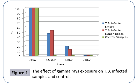
In the present study, all collected samples from T.B. infected carcasses were exposed to Gamma radiation at three absorbed dose levels of 2.5, 5 and 7.5 kGy, showed that at 0.0 kGy all samples still 100% infected and 46.6% still infected at 2.5 kGy and 20% still infected at 5 kGy and, At 7.5% all examined samples are failed to be detected of T.B. Infected offal's. Moreover, the examined samples of T.B. Infected lymph nodes showed that at 0 kGy all samples still 100% infected and 53.3% still infected at 2.5 kGy and 13.3% still infected at 5 kGy and, at 7.5% all examined samples are free from mycobacterium infection (Table 1).
Table 1 The effect of gamma rays exposureon T.B. infected samples and control.
| Tissue | No. of samples | Used doses | |||
|---|---|---|---|---|---|
| 0 | 2.5 | 5 | 7.5 | ||
| PCR Positive | |||||
| T.B.Infected offal's (liverandkidney) |
15 | 15 | 7 | 3 | 0 |
| T.B.Infected Lymph nodes (Hepatic and Renal) |
15 | 15 | 8 | 2 | 0 |
| Control Samples | 5 | 5 | --- | --- | --- |
As mentioned in a Table 1 the infected samples with Mycobacterium tuberculosis bovine type which exposure to doses levels 2.5, 5 and 7.5 kGy which were re-examined for detection of Mycobacterium tuberculosis bovine type by using RT-PCR showed that, the T.B. infected offal's, the PCR can confirmed the infection of 7 samples out of 15 tested samples at 2.5 kGy while 3 positive samples only at 5 kGy and failed to be detected at 7.5 kGy.
Also, the T.B. infected lymph nodes, the PCR can confirmed the infection of 8 samples out of 15 tested samples at 2.5 kGy while 2 positive samples only at 5 kGy and failed to be detected at 7.5 kGy (Figure 1).
Foodborne pathogens are associated with food processing plants and slaughtered animals, the basic raw materials of the food industry. With the exception of foods that are thermally treated to the degree of sterilization, all food products are frequently associated with microorganisms [32]. The most important risk of these infectious diseases is tuberculosis Bovine. Tuberculosis is now generally perceived to represent the greatest threat to cattle health, its caused by M.bovis and can affect a large number of species, including humans [33].
Ionizing radiation was first patented in 1905 as a microbial inactivation technology and was first evaluated in 1921 against trichinae in pork [34]. Ionizing radiation has enough energy to remove electrons from atoms and leads to ion formation. Ionizing radiation comes in different forms depending on the source (X-ray, gamma rays, and beta rays); however, all forms exert their effects by “stripping” electrons from atoms. This irradiation causes breaks in the DNA and/or RNA helix and leads to the disruption of normal cellular functions by damaging nucleic acids by either direct or indirect effect [35].
Food irradiation processing technology and gamma irradiation are widely used as a safe and proven method worldwide for the preservation of food products. Food irradiation was approved by the FAO/IAEA/WHO in 1981 with maximum irradiation doses of up to 10 kGy [36]. More than 50 countries worldwide have approved the irradiation of over 60 food products [37,38]. Furthermore, government regulation considerably varies from country to country. Each country has adopted its own unique approach to the introduction, approval, and regulation of this technology in food production. Although there is agreement among international committee experts that food is safe and wholesome for consumption after irradiation with doses up to 10 kGy, food irradiation at this dose has not been approved in any country. Most countries approve food irradiation on a case-bycase basis [39] (Table 2).
Table 2 The effect of exposure of gamma rays on T.B. infected samples.
| Tissue | No. of samples | Used doses | |||
|---|---|---|---|---|---|
| 0 | 2.5 | 5 | 7.5 | ||
| Reaction rate % | |||||
| T.B.Infected offal's (liverandkidney) |
15 | 100 % | 46.6% | 20 % | 0 % |
| T.B.Infected Lymph nodes (Hepatic and Renal) |
15 | 100 % | 53.3 % | 13.3 % | 0 % |
As mentioned in Table 2 the conclusion for T.B. Infected samples the reduction rate is decreased by increase the dose level of Gamma rays. At 0 kGy all samples still 100% infected and 46.6% still infected at 2.5 kGy and 20% still infected at 5 kGy and, At 7.5% all examined samples are free regarding samples of T.B. Infected offal's. And also, the examined samples of T.B. Infected lymph nodes showed that at 0 kGy all samples still 100% infected and 53.3% still infected at 2.5 kGy and 13.3% still infected at 5 kGy and, at 7.5% all examined samples are free from mycobacterium infection (Table 3).
Table 3 The effect on (color, odor and texture) parameter after exposure to gamma rays on T.B. infected samples.
| Parameter | No. of samples | SCORE | |||||
|---|---|---|---|---|---|---|---|
| 24 hours | 1 week | ||||||
| 1 | 2 | 3 | 1 | 2 | 3 | ||
| Color | 25 | 5 | 20 | --- | 25 | --- | --- |
| Odor | 25 | --- | 25 | --- | 25 | --- | --- |
| Texture | 25 | 20 | 5 | --- | 20 | 5 | --- |
Note: 1. No. of samples with normal (Color, Odor and Texture);2.No. of samples with slight changes in (Color, Odor and Texture); 3. No. of samples with abnormal changes in (Color, Odor and Texture)
Conclusion
The obtained results from our study about the effect on (Color and Odor and Texture) parameter after exposure to Gamma rays on T.B. infected samples proved that most of tested samples have slight changes in color (pale color), odor (characteristic odor of irradiation) and texture (friable) in the first 24 hours and all tested samples have been returned back into the normal parameter after 1 week.
More investigations were required for the application of Gamma rays to eliminate the tubercle bacilli from the infected offal's to save the huge amount of contamination meat even to use it as low grade meat. Further, work is needed to evaluate the in vivo assays after feeding the experimental animals on the irradiated food stuff.
References
- Center for Disease Control (2004) Preliminary FoodNet data on the incidence of infection with pathogens transmitted commonly through food-Selected sites, United States, 2003. MMWR Morb Mortal Wkly Rep 53: 338-343.
- USDA/ERS (2000) Economics of foodborne disease: Feature. United States Department of Agriculture, Economic Research Service, Washington, D.C.
- USDA/FSIS (2004) FSIS Recalls: Closed federal cases. United States Department of Agriculture, Food Safety and Inspection Service, Washington, D.C.
- Lagunas-Solar MC (1995) Radiation processing of foods: An overview of scientific principles and current status. J Food Prot 58: 186-192.
- Morehouse KM (2002) Food irradiation-US regulatory considerations. Radiat Phys Chem 63: 281-284.
- Oliveira IB, Sabato SF (2002) Brazilian consumer acceptance of irradiated food: initial trials. The Americas Nuclear Energy Symposiums.
- Resurreccion AVA, Galvez FCF (1999) Will consumer buy irradiated beef? Food Tech 53: 52-55.
- Hines ME, Kreeger JM, Herron AJ (1995) Mycobacterial infections of animals: Pathology and Pathogenesis. Lab Anim Sci 45: 334-351.
- Martin SW, Dietrich RA, Genno P, Heuschele WP, Jones RL, et al. (1994) Livestock disease eradication: Evaluation of the cooperative state federal bovine tuberculosis eradication program. National Academy of Sciences, Washington D.C,pp: 1-97.
- Samad MA (2000) Animal husbandry and veterinary science. BAU Campus, Mymensingh, Bangladesh.
- Radostis OM, Gay CC, Peter DC, Hinchdiff KW (2000) Veterinary Medicine-A textbook of the diseases of cattle, horses, sheep, pigs and goats (10thedn.). Saunders, USA.
- Prasad HK, Singhal A, Mishra A, Shah NP, Katoch VM, et al. (2005) Bovine tuberculosis in India: Potential basis for zoonosis. Tuberculosis 85: 421-428.
- Bartzatt R, Cirillo SLG, Cirillo JD (2008) Determination of molecular properties effectuating the growth inhibition of mycobacterium tuberculosis by various small molecule hydrazides.Lett Drug Des Discov 5: 162-168.
- JunqueiraGoncalves MP, Galotto MJ, Valenzuela X, Dinten CM, Aguirre P, et al. (2011) Perception and view of consumers on food irradiation and the Radura symbol. Radiation Physics &Chemistry 80: 119-122.
- Fox JA (2002) Influences on purchase of irradiated foods. Food Technol 56: 34-37.
- Landgraf M, Gaularte L, Martins C, Cestari A, Nunes T, et al. (2006) Use of irradiation to improve the microbiological safety of minimally processed fruits and vegetables. IAEA-TECDOC 1530: 41-59.
- Marcotte M (2005) Effect of irradiation on spices, herbs, and seasonings-comparison with ethylene oxide fumigation.
- Davis KJ, Sebranek J, Huff-Lonergan E, Ahn DU, Lonergan SM (2004) The effects of irradiation on quality of injected fresh pork loins. Meat Sci 67: 395-401.
- Dogbevi MK, Vachon C, Lacroix M (1999) Physicochemical and microbiological changes in irradiated fresh pork loins. Meat Sci 51: 349-54.
- Borsa J, Lacroix M, Ouattara B, Chiasson F (2004) Radiosensitization: Enhancing the radiation inactivation of food borne bacteria. Radiat Phys Chem 71: 137-41.
- Unluturk S, Atilgan MR, Baysal AH, Tari C (2007) Use of UV-C radiation as a non-thermal process for liquid egg products (LEP). J Food Eng 85: 561-68.
- Byrd-Bredbenner C, Cohn MN, Farber JM, Harris LJ, Roberts T, et al. (2015) Food safety considerations for innovative nutrition solutions. Ann N Y Acad Sci1347: 29-44.
- Khan I, Oh DH (2016) Integration of nisin into nanoparticles for application in foods. Innov Food Sci Emerg Technol 34: 376-384.
- Khan I, Ullah S, Oh DH (2016) Chitosan grafted monomethylfumaric acid as a potential food preservative. Carbohydr Polym 152: 87-96.
- Marks J (1972) Ending the routine guinea pig test. Tubercle 53: 31-34.
- Kubica GP (1973) Differential identification of Mycobacteria. VII.key features for identification of clinically significant mycobacteria. Am Rev Respir Dis 107:9-21.
- Wards BJ, Collins DM, De-Lisle GW (1995) Detection of M. bovis in tissues by polymerase chain reaction. Vet Microbio l43: 227-240.
- Xiu-yun J, Wang C, Zhang P, Zhao-yang H (2006) Cloning and Expression of Mycobacterium tuberculosis bovine type Secreted Protein MPB83 in Escherichia coli. Biochem Mol Biol 39: 22-25.
- Ben KI, Boschiroli ML, Souissi F, Cherif N, Benzarti M, et al. (2011) Isolation and molecular characterization of Mycobacterium tuberculosisbovine type from raw milk in Tunisia. Afr Health Sci 11: 52-55.
- vanSoolingen D, Hermans PW, de Haas PE, Soll DR, van Embden JD (1991) Occurrence and Stability of Insertion Sequences in Mycobacterium tuberculosis Complex Strains: Evaluation of an Insertion Sequence-Dependent DNA Polymorphism as a Tool in the Epidemiology of Tuberculosis. J Clin Microbiol 29: 2578-2586.
- Miller RB (2005) Food irradiation using electron beams. In: Miller RB (ed.), Minimally Processed Foods. CRC Press, Boca Raton, FL. pp: 279-300.
- Pattanayaiying R, H-Kittikun A, Cutter CN (2015) Incorporation of nisin Z and lauricarginate into pullulan films to inhibit foodborne pathogens associated with fresh and ready-to-eat muscle foods. Int J Food Microbiol 207:77-82.
- Cobner A (2003) Bovine tuberculosis: clinical update and on-farm advice. In Practice 25: 606-613.
- Negut CD, Bercu V, Duliu OG (2012) Defects induced by gamma irradiation in historical pigments. J Cult Herit 13: 397-403.
- Kuan YH, Bhat R, Patras A, Karim AA (2013) Radiation processing of food proteins-A review on the recent developments. Trends Food Sci Technol 30: 105-120.
- Lacroix M, Ouattara B (2000) Combined industrial processes with irradiation to assure innocuity and preservation of food products-A review. Food Res Int 33:719-724.
- Eustice RF (2015) Food-Irradiation.
- World Health Organisation (2015) Food-Safety. WHO, Geneva.
- Food and Drug Administration (2016) U.S. regulatory requirements for irradiating foods.

Open Access Journals
- Aquaculture & Veterinary Science
- Chemistry & Chemical Sciences
- Clinical Sciences
- Engineering
- General Science
- Genetics & Molecular Biology
- Health Care & Nursing
- Immunology & Microbiology
- Materials Science
- Mathematics & Physics
- Medical Sciences
- Neurology & Psychiatry
- Oncology & Cancer Science
- Pharmaceutical Sciences

