Statistical Analysis of Mobile Radiation and Impact on Health: A Case Study on Occurrence of Brain Tumors
1Department of CSE, Geethanjali College of Engineering and Technology, India
2Defence Research and Development Organization, Hyderabad, India
- *Corresponding Author:
- Prisilla Jayanthi
Department of CSE, Geethanjali College of Engineering and Technology
Cheeryala, India
Tel: +919502952210
E-mail: prisillaj28@gmail.com
Received date: November 01, 2018; Accepted date: November 20, 2018; Published date: November 30, 2018
Citation: Jayanthi P, Krishna IVM (2018) Statistical Analysis of Mobile Radiation and Impact on Health: A Case Study on Occurrence of Brain Tumors. J Health Hyg. Vol.2 No:1:2
Copyright: © 2018 Jayanthi P, et al. This is an open-access article distributed under the terms of the Creative Commons Attribution License, which permits unrestricted use, distribution, and reproduction in any medium, provided the original author and source are credited.
Abstract
To analyze whether mobile phone radiation is one of the source for brain tumor. This study is carried out by odd ratios.
The mobile radiation seems to be severe among children than the adults because their skulls are tiny and thinner. Mobile phones emit radiations and RF-EMF is considered as a Group 2B (carcinogen) and the survey suggests that 10-year latency period is practically enough for the development of tumors. The studies from various groups involve cases of 35487 and controls about 82609. The results for meta- analysis of glioma gave an odd ratio OR=1.10, 95% CI=0.79-1.54 and for the latency period ≥ 10 years, OR=1.38, 95% CI=0.70-2.73. The highest risk was found in the age group below 20 years from the Hardell Group.
Keywords
Exposure; Mobiles; Radiation; Risk
Introduction
Intense use of wireless communication has raised a concern over the human health, an increased risk for brain tumors. Mobile phones and cordless phones emit radiofrequency (RF) radiation when in use. The minimum period was ≥ 10 years or ≥ 1640 hours for the development of tumors using wireless phones with the higher the exposure on the same side of the brain (ipsilateral) whereas the contralateral side is exposed less [1].
In May 2011, an approach for the scientific evidence of the risk for brain tumor was completed by the International Agency for Research on Cancer (IARC) at the World Health Organization (WHO). IARC is independently funded and has categorized the extremely low frequency (ELF) as a Group 2B carcinogen.
The radio frequency energy emitted by mobile phones can be absorbed by brain tissues near to the mobile; is a form of nonionizing electromagnetic radiation. The brand of mobile phone, the distance between the phone and user and user’s distance from cell phone towers are the key factors in the amount of radio frequency energy a mobile user is exposed. In 1998, the International Commission on Non-Ionizing Radiation Protection (ICNIRP) has established the exposure guidelines and short-term thermal effect from RF radiation [2].
Child’s Brain Endanger
A child is in danger when the child holds the mobile phone, Ipad or any radiating device, as a child’s brain can absorb higher rates than adults due to the smaller head size, thinner skull bones and higher brain conductivity. The study reveals that most of the children below two years are left with mobiles, i-pad or any other electronic gadget because the parents are busy in their jobs and regular routine work. Henceforth, children are at higher risk of exposure to carcinogens than adults, and the smaller the child the greater the risk [3,4]. There are several studies that prove that children absorb more Micro Wave Radiation (MWR) than adults. In 1996, one of the study concluded that the penetration of absorbed MWR was deeper into the children’s brain of age 5 to 10 years [5,6]. In 2008, a French senior researcher named Joe Wiart, pointed that the child’s brain tissue absorbed two times more MWR than adults [7]. In 2009, the study stated that the Central Nervous System absorption in children’s is higher (~2x) as the MWR source is closer and skin and bone layers are thinner and bone marrow exposure (~10x) varies with the age [8]. In 2010, Andreas Christ and his team described that the child’s hippocampus and hypothalamus absorbs 1.6–3.1 times greater and the cerebellum absorbs 2.5 times higher MWR compared to adults; the bone marrow of child absorbs 10 times more MWR radiation than adults [9,10].
Mobile Phones SAR Evaluation
The Specific Absorption Rate (SAR; W kg-1) depends on the type of phone used; one can calculate the SAR per number of hours exposed [11]. Based on the type of phone manufacturer, few phones give peak SAR value above the ear, some beneath the ear and few even below the ear. The SAR value will affect the human ear and later damage the human brain. And whose ears are affected with SAR cannot hear properly. Jonna Wilen has calculated mean values of SAR1g for a certain number of people who had a particular symptom (W–S) and those who do not have the symptom (WO–S). Jonna’s study from her observation concluded that 30 out of 44 comparisons (W–Smean/WO–Smean) are greater than 1. The symptom discomfort is known from W– S=95, SAR1g=0.67 (0.23) and WO–S=2094, SAR1g=0.59 (0.27), (W–Smean / WO–Smean) =1.14. The symptom concentration W– S=180, SAR1g=0.65 (0.25) and WO–S=2009, SAR1g=0.59 (0.27) and (W–Smean / WO–Smean)=1.10 below the ear (volume 3), the equivalent differences were larger than 10% [12,13]. The initial increase for brain tumor risk associated with mobile phones was issued before 10 years and was found for ipsilateral mobile phone use [14,15].
Statistical Methodology
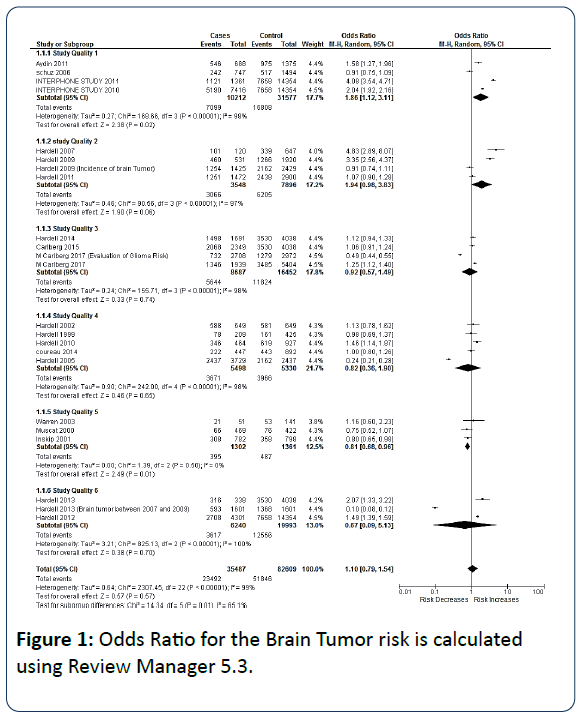
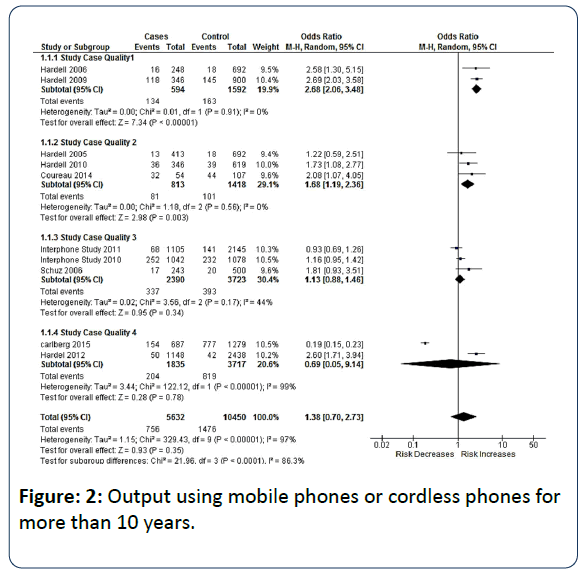
For data analysis, odds ratio was calculated using Review Manager 5.3. The random effects model was used to measure summary odds ratio, based on chi-square test, tau squared and I-squared statistics for heterogeneity as shown in Figures 1 and 2 [16-19]. Latency is defined as the first year of use of wireless to the diagnosis year. Latency was analyzed using periods groups >10 years, 10-15 years, 15-20 years and >25 years.
Figure 1: Odds Ratio for the Brain Tumor risk is calculated using Review Manager 5.3.
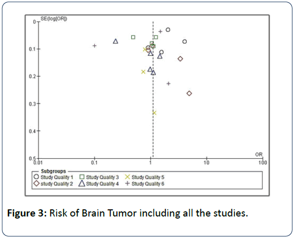
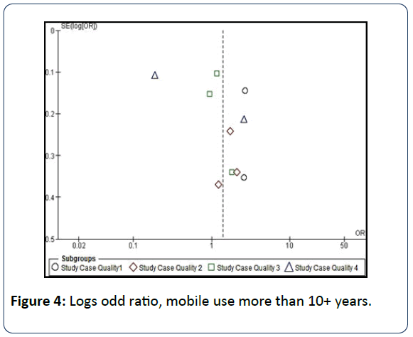
Based on Hardell group and Interphone group studies the meta-analyses were performed with the mobile phones use. The model was chosen based on the latency and the number of hours (≥ 10 years and ≥ 1640 hrs) a mobile is used to test for heterogeneity. The total number cases 35487 and control 82609 evaluated for all 23 studies considered from the Hardell group and Interphone study group in the analysis of patients with glioma yielded OR=1.10 95% CI=0.79-1.54 (p=0.57). The relationship between the study qualities and odd ratios are shown in graphs in Figures 3 and 4.
Figure 3: Risk of Brain Tumor including all the studies.
Odd Ratios for Mobile Use Over 10+ Years
Lonn et al. and group, Karolinska Institute in Sweden conducted a study on glioma and meningioma cases. The mobile use of ≥ 10 years showed for ipsilateral glioma OR=1.6, 95% CI=0.8-3.4 for 15 cases and for contralateral glioma OR=0.7 95% CI=0.3-1.5 for 11 cases. Shoemaker et al. of the interphone study showed the results for acoustic neuroma from six regions, the results for 678 cases of lifetime use (≥ 10 years) showed OR=1.8, 95% CI=1.1-3.1 for ipsilateral acoustic neuroma, and OR=0.9 95% CI=0.5-1.8 for contralateral tumor. The Danish of the interphone study for ≥ 10 years produced OR=1.0 (0.3 to 3.2) for meningioma, low–grade glioma OR=0.5, 95% CI=0.2-1.3 and high-grade glioma OR=0.5, 95% CI=0.2-1.3 for 252 glioma cases, 175 meningioma cases with 822 controls. Hepworth et al. England showed results on glioma as a part of the interphone study for ≥ 10 years, ipsilateral provided OR=1.6, 95% CI=0.9-2.8 and contralateral OR=0.8, 95% CI=0.4-1.4 for 966 cases [1]. The interphone study group pointed that the time a mobile used for ≥ 10 years OR=0.83, 95% CI=0.58–1.19 for 10 years after regular use and for the hours ≥ 1640 h of cumulative call time it was 2.79 (1.51–5.16) [20,21]. Hardell, the study on the cumulative use of wireless phones in different quartiles, the quartile >1486 h gave OR=2.6, 95% CI=1.5-4.4 for mobile phone and wireless phone use produced OR=2.2, 95% CI=1.5-3.4 [22]. Hardell, analyzed that using a cordless phone gave an increased highest risk in diagnosing of glioma in the latency group >10 years OR=3.8, 95% CI=1.8-8.1 and for glioma with the mobile use ≥ 1640 hrs; the increased risk in the temporal lobe giving OR=1.87, 95% CI=1.09-3.22 from Table 1 [23].
| Study | Cases/events | OR 95%CI | Comments |
|---|---|---|---|
| Hardell (1997-2003) | 148/57 | 2.9(1.8-4.7) | Latency period >10 years, mobile phone (ipsilateral) |
| 148/20 | 3.8(1.8-8.1) | Latency period >10 years, cordless phone (ipsilateral) | |
| Interphone study group (2000- 2004) | 2708/210 | .40(1.03-1.89) | Mobile phone Cumulative hours ≥ 1640 hrs |
| 2708/78 | .87(1.09-3.22) | Mobile phone cumulative Hours ≥ 1640 hrs, tumor in the temporal lobe |
|
| 2708/100 | .96(1.22-3.16) | Ipsilateral phone usage cumulative hours ≥ 1640h hrs |
Table 1: Summary of studies of wireless phones usage and glioma risk.
Carlberg concluded that the longer latency of 10+ years OR=1.62, 95% CI=1.20–2.19. The three studies revealed a consistent increase of glioma risk with latency. The highest OR with longest latency of 10+ years with 732 exposed cases and 1,279 exposed controls [24]. Carlberg analyzed that the cumulative use >3358 hours relates to >90th percentile. An increased risk for mobile phone (2G and 3G) with OR=1.5, 95% CI=1.0005-2.3(p-trend=0.045) and cordless phone showed OR=2.1, 95% CI=0.7-6.3 (p-trend=0.65) [25].
Hardell, the peak cumulative use of mobile phones ≥ 1640 h for glioma OR=1.40 95% CI=1.03-1.89 was calculated. The risk increased for ipsilateral use OR=1.96, 95% CI=1.22-3.16. In temporal lobe, the highest risk was found with more exposure in the anatomical area [26].
Findings
Carlberg examined a largest case-control study on brain tumors and occupational exposure to ELF-EMF. The interocc study proved a relation between exposure to ELF-EMF and glioma and finally concluded that the final phases of astrocytoma grade IV for occupational ELF-EMF exposure had an increased risk [18]. Carlberg concluded a fact that the occupational ELF-EMF exposure has an increased risk of glioma. Bradford Hill’s viewpoints on association on RF radiation and glioma risk. Hence concluded that glioma is caused by RF radiation [24].
Hardell, the questionnaire was answered by 1498 of 1691 cases, of whom 879 were men and 619 women, of 4038 controls, 1492 men and 2038 women participated to give the total of 3530. The glioma risk at various age groups for the wireless phone use was found to be increasing. The risk increased in both mobile and cordless phones and OR is high before the age of 20 years. Children are more exposed to RFEMF than adults as higher conductivity in the brain tissue and a smaller head [17]. Hardell concluded that the volume of tumor increased for ipsilateral use of mobile phones of the digital 2G type and for cordless phones per year of latency. Interphone Study, the glioma cases diagnosed were 23% of 2708 cases before the age of 40.
Hardell suggested that a consistent association between use of mobile or cordless phones and astrocytoma grade I-IV and acoustic neuroma. For latency period >10 years, the risk was highest for ipsilateral exposure to microwaves. The greater risk for persons who started to use mobiles before the age of 20 years were identified in Sweden during 2000-2007 and the results were supported by the increase of incidence of astrocytoma [27]. Hardell revealed a non –significant increase risk for brain tumors located in the temporal or occipital lobe was identified for those who used cell phones on the same side of the head. Acoustic neuroma develops with higher exposure to microwave radiation from a mobile phone [28].
Comparative Analysis
The comparison was done using the results obtained by the calculation carried out using review manager and the respective group results as follows:
Inskip, reported that the relative risk of gliomas RR=0.9, 95% CI=0.7-1.1 with the cases=308 and controls=358. According to Figure 1, Inskip, the relative risk RR=0.88, 95% CI=0.78- 0.99 data was not shown in the Figure [21].
Hardell, the evaluated results of glioma (n=1380) and use of mobile and cordless phones in different latency group as >10 years, >15-20 years, >20-25 years, and >25 years. The cordless phone use gave OR=1.65, 95% CI=1.32-2.05 in the latency group >15-20 years. The digital type 2G, 3G and cordless showed OR=1.54, 95% CI=1.34-1.78 in the latency group >10 years for wireless phones. The OR=1.35, 95% CI=0.51-3.58 for cordless for latency >20-25 years. The increased risk for wireless phones is given by OR=1.032, 95% CI=1.019-1.046 [17].
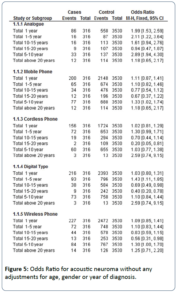
Hardell pointed that the OR=0.81, 95% CI=0.70-0.94 for mobile phone use >1 year for glioma and further risk increased for glioma in the temporal lobe yielding OR=1.87, 95% CI=1.09-3.22, the results from Figure 1 reflects OR=1.49, 95% CI=1.39-1.59. The regular use of mobile phone and cordless phone for latency period >10 years yielded OR=2.9, 95% CI=1.8-4.7 for ipsilateral use. It was found doubling of glioma risk for total wireless phone use with OR=2.1, 95% CI=1.6-2.8. The results from review manager showed OR=2.60, 95% CI=1.71-3.94 for more than 10 years [23]. Hardell showed for acoustics neuroma over the latency period >10 years OR=1.3, 95% CI=0.6-2.8 whereas from Figure 1, the obtained OR=4.83, 95% CI=2.89-8.07 [1]. The result of mobile phones, cordless phone and wireless phone with the OR result analysis for 316 cases and 3530 controls by Hardell. From Figure 5, mobile phone use for 5 to 10 years OR=1.33 95% CI=1.02-1.74, cordless phone OR=1.03, 95% CI=0.77-1.38 and wireless phone use shows OR=1.30, 95% CI=1.00-1.70.
Hardell group has shown with an adjustment of age, gender and year of diagnosis and the latency >5-10 years the OR=2.3, 95% CI=1.6-3.3 for mobile phone, for cordless phone OR=1.6 95% CI=1.1-2.5 and wireless phone OR=1.9, 95% CI=1.3-2.7 for the same number of cases and controls.
The output of odds ratio using Review Manager 5.3 evaluated for all the studies for the total number of cases 35487 and control 82609 from the Hardell group and Interphone study group for the analysis of patients with glioma yielded OR =1.10, 95% CI=0.79-1.54 (p=0.57) which on comparing to the odds ratio OR=1.03, 95% CI=0.92-1.14 (p=0.64) calculated by the group from AIIMS (Delhi) using Review Manager with total cases 12426 and controls 19334. It indicates significant increase of OR=0.07.
Conclusion
Results from the above various case- control studies on brain tumors and mobile phone usage for above 10 years suggests that there is an increased risk in glioma. The adults are to be aware when their children are left with mobile or any electronic gadget for entertainment, that there is risk for the child’s health. The overall result of meta-analysis showed a significant increase of 1.38 times OR in brain tumor risk.
Acknowledgements
I am thankful for Hardell group, Devra Lee Davis Group, and Dr. Jonna Wilen for their extended assistance of sharing their research papers promptly and my sincere thanks to Mr. Michael Carlberg for his on-time and constant support rendered to me. My special thanks for Dr. Kameshwar Prasad, Chief Neurologist, AIIMS for spending his valuable for discussion. All the data is included with the permission of all the concerned authors from different articles, published by the same group of authors.
Conflicts of Interest
The authors declare that there are no conflicts of interest regarding the publication of this paper.
References
- Hardell L, Carlberg M, Söderqvist M, Mild KH, Morgan LL, et al. (2007) Long-term use of cellular phones and brain tumours: increased risk associated with use for > 10 years. Occup Environ Med 64: 626–632.
- Hardell L (2017) World Health Organization, radiofrequency radiation and health - a hard nut to crack (Review). Int J Oncol 51: 405-413.
- Hardell L, Carlberg M, Mild KH (2005) Pooled analysis of two case-control studies on the use of cellular and cordless telephones and risk of benign tumors diagnosed during 1997-2003. Int Arch Occup Environ Health 28: 509-518.
- Morgana LL (2014) Why children absorb more microwave radiation than adults: The consequences. Journal of Microscopy and Ultrastructure 2: 197–204.
- Aydin D, Feychting M, Schüz J, Tynes T, Andersen TV, et al. (2011) Mobile Phone Use and Brain Tumors in Children and Adolescents: A Multicenter Case–Control Study. J Natl Cancer Inst 103: 1264-1276.
- Gandhi OP, Lazzi G, Furse CM (1996) Electromagnetic absorption in the human head and neck for mobile telephones at 835 and 1900 MHz?. IEEE Trans Micro Theory Tech 44: 1884-1897.
- Wiart J, Hadjem A, Wong MF, Bloch I (2008) Analysis of RF exposure in the head tissues of children and adults. Phys Med Biol 53; 3681-3695
- Kuster N (2009) Past, current, and future research on the exposure of children. Foundation for Research on Information Technology in Society (IT’IS). Foundation Internal Report
- Christ A, Gosselin MC, Christopoulou M, Kühn S, Kuster N (2010) Age-dependent tissue- specific exposure of cell phone users. Phys Med Biol 55: 1767-1783.
- Gandhi OP, Morgan LL, de Salles AA, Han YY, Herberman RB, et al. (2012) Exposure Limits: The underestimation of absorbed cell phone radiation, especially in children. Electromagn Biol Med 31: 34-51.
- Hardell L, Mild KH, Carlberg M (2002) Case-control study on the use of cellular and cordless phones and the risk for malignant brain tumours. Int J Radiat Biol 78: 931- 936.
- Wilén J, Sandström M, Mild KH (2003) Subjective symptoms among mobile phone users - a consequence of absorption of radiofrequency fields? Bioelectromagnetics 24: 152-159.
- Hardell L, Mild KH, Påhlson A, Hallquist A (2001) Ionizing radiation, cellular telephones and the risk for brain tumours”. Eur J Cancer Prev 10: 523-529,
- Cardis E, Deltour I, Mann S, Moissonnier M, Taki M, et al. (2008) Distribution of RF energy emitted by mobile phones in anatomical structures of the brain. Phys Med Biol 53: 2771-2783.
- Hardell L, Carlberg M, Söderqvist F, Mild KH (2013) Case- control study of the association between malignant brain tumours diagnosed between 2007 and 2009 and mobile and cordless phone use. Int J Oncol 43: 1833-1845.
- The Interphone Study Group (2010) Brain tumour risk in relation to mobile telephone use: Results of the Interphone international case-control study. Int J Epidemiol 39: 675-694.
- Hardell L, Carlberg M (2014) Mobile phone and cordless phone use and the risk for glioma –Analysis of pooled case-control studies in Sweden, 1997-2003 and 2007-2009. Pathophysiol 22: 1–13.
- Carlberg M, Koppel T, Ahonen M, Hardell L (2017) Case-control study on occupational exposure to extremely low-frequency electromagnetic fields and glioma risk. Am J Ind Med 60: 494-503.
- Prasad M, Kathuria P, Nair P, Kumar A, Prasad K (2017) Mobile phone use and risk of brain tumors: a systematic review of the association between study quality, source of funding, and research outcomes. Neurol Sci 38: 797-810.
- The INTERPHONE Study Group (2011) Acoustic neuroma risk in relation to mobile telephone use: Results of the INTERPHONE international case–control study. Cancer Epidemiol 35: 453-464.
- Inskip PD, Tarone RE, Hatch EE, Wilcosky TC, Shapiro WR (2001) Cellular-Telephone Use And Brain Tumors. N Engl J Med 344: 79-86.
- Hardell L, Carlberg M, Söderqvist F, Mild KH (2013) Pooled analysis of case-control studies on acoustic neuroma diagnosed 1997-2003 and 2007- 2009 and use of mobile and cordless phones. Int J Oncol 43: 1036-1044.
- Hardell L, Carlberg M, Mild KH (2013) Use of mobile phones and cordless phones is associated with increased risk for glioma and acoustic neuroma. Pathophysiology 20; 85-110.
- Carlberg M, Hardell L (2017) Evaluation of Mobile Phone and Cordless Phone Use and Glioma Risk Using the Bradford Hill Viewpoints from 1965 on the Association or Causation. Biomed Res Int 2017:9218486.
- Carlberg M, Hardell L (2015) Pooled analysis of Swedish case-control studies during 1997-2003 and 2007-2009 on meningioma risk associated with the use of mobile and cordless phones. Oncol Rep 33: 3093-3098.
- Hardell L, Carlberg M, Mild KM (2011) Pooled analysis of case- control studies on malignant brain tumors and the use of mobile and cordless phones, including living and deceased subjects. Int J Oncol 38: 1465-1474,
- Hardell L, Carlberg M (2009) Mobile phones, cordless phones and the risk for brain tumors. Int J Oncol.
- Hardell L, Näsman A, Påhlson A, Hallquist A, Mild KH (1999) Use of cellular telephones and risk of brain tumors: A case- control study. Int J Oncol 15: 113-116.

Open Access Journals
- Aquaculture & Veterinary Science
- Chemistry & Chemical Sciences
- Clinical Sciences
- Engineering
- General Science
- Genetics & Molecular Biology
- Health Care & Nursing
- Immunology & Microbiology
- Materials Science
- Mathematics & Physics
- Medical Sciences
- Neurology & Psychiatry
- Oncology & Cancer Science
- Pharmaceutical Sciences
