Rosai-Dorfman Disease: A clinical Case With Systemic Nodal Presentation and Literature Review
Enoch Alvarez Rodriguez, Cynthia Shanat Cruz Medina*, Eduardo Tellez Bernal, Miriam Ixel Escamilla Lopez, Andrea Castro Sanchez, Salvador Macias Diaz, Maria de Jesus Gonzalez Blanco, Jose Maria Ortega Solar, Alejandro Esquivel Lu, Fernanda Alicia Baldeon Figueroa and Maria Fernanda Burgos Vazquez, Valeria Mendoza Ortiz
Department of Oncology Pediatric, University of Benemerita Universidad Autonoma de Puebla, Puebla, Mexico
- *Corresponding Author:
- Cynthia Shanat Cruz Medina
Department of Oncology Pediatric,
University of Benemerita Universidad Autonoma de Puebla,
Puebla,
Mexico
Tel: 4432792889
E-mail: ped_hope@hotmail.com
Received date: September 26, 2023, Manuscript No. IPMO-23-17945; Editor assigned date: September 29, 2023, PreQC No. IPMO-23-17945 (PQ); Reviewed date: October 13, 2023, QC No. IPMO-23-17945; Revised date: December 27, 2023, Manuscript No. IPMO-23-17945 (R); Published date: Janyary 03, 2024, DOI: 10.36648/IPMO.7.1.001
Citation: Medina CSC, Rodriguez EA, Bernal ET, Lopez EI, Sanchez AC, et al. ( 2024) Rosai-Dorfman Disease: A Clinical Case with Systemic Nodal Presentation and Literature Review. J Med Oncol Vol:7 No:1
Abstract
Rosai-Dorfman disease, also known as sinus histiocytosis with massive lymphadenopathy is described as a benign, rare and self-limiting clinicopathologic entity characterized by histiocytic proliferation and painless lymphadenopathy. We report an unusual presentation where the condition occurs both at nodal and systemic presentation.
Rosai-Dorfman; Case report; Systemic nodal manifestation; Sinus histiocytosis; Massive lymphadenopathy; Extranodal
Introduction
Destombes Rosai-Dorfman Disease (RDD) was first described in 1965 by Dr. Paul Destombes, french pathologist, who first observed voluminous cervical adenopathy that when studied showed an accumulation of histiocytes with abundant and pale cytoplasm and fibrous capsule presence in 1969, John Rosai and Ronald Dorfman describe it as a rare, benign and self-limited cline-pathologic entity, also called histiocytosis with massive lymphadenopathy, that presents itself as a painless and bilateral massive adenopathy. It tends to affect young patients and has a nodal presentation (classical), extra-nodal and cutaneous [1].
Case Presentation
17-year-old female patient, from Teloloapan, with surgical history at 3 years old in the hospital of Guerrero for excision of tumorous left forearm, of which there is no pathological report.
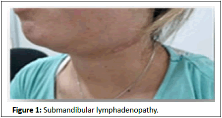
Current illness starts in December 2020 with presence of sub-mandibular adenopathy with a progressive size growth with 2 months evolution, without any other aggregate symptoms, with a 7 days fever of un-quantified evolution, without strong schedule (Figure 1).
In the neck USG you can observe multiple adenopathy in all ganglionic levels of atypical characteristics with augmented vascularity and necrotic zones, of which the largest are located at level VI right 9 × 10 mm and left 6 × 7 mm.
The submandibular adenopathy diagnostic is given in a 2 months study of the evolution with a diagnosis of sinusoidal hyperplasia with histiocytosis, which was diagnosed in December 2020. The USG from October 2021 show a presence of right cervical lymph nodes in segments II, II and IV, A and B, left segments II and III up to 5 cm diameter, on the left forearm, in the internal lateral face of 2 cm diameter, refers to radicular type pain by compression, reflected in right hemichalea hemicrania, pulsing of 6/10 intensity with gradual intensity increase.
The simple and contrasted thorax and abdomen study is made, in the neck you can observe a homogenous thyroid gland, the trachea is centered.
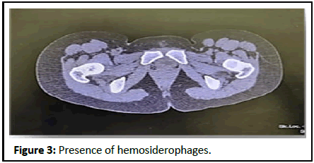
In the simple head CT-scan you can observe cervical lymph nodes of inflammatory characteristics, the most representative being bilateral in level IIB, being VB the biggest at 17 × 12 mm in axial cut. Also, there is a presence of inflammatory nodes in inguinal zone, axillar zone and mesenteric nodes.
A dose of 200 mg of cyclophosphamide is prescribed, followed by steroids and 20 mg of methotrexate every week, an appointment is given in 15 days with a study of head imaging for the new lesion of the right temporal and malar region.
The simple head CT-scan is made where you can observe cervical lymph nodes of inflammatory characteristics, the most representative being bilateral in level IIB, being VB the biggest at 17 × 12 mm in axial cut (Figure 2).
Figure 2: Scan of the simple skull is performed where cervical lymph nodes with inflammatory characteristics are observed, but preserving their morphology, the most representative at bilateral level IIB, in addition at level VB the largest node is observed with a measurement of 17 × 12 mm in the axial section. The trachea is observed central, in its lumen at the supracarinal level an ovoid image is observed, with lobulated edges, adhered to the left lateral wall, hypodense with significant enhancement and measures 9 × 4 mm with a probable relationship to mucus, mesenteric ganglia with an increase in their size. Number and diameter, predominantly pericecal and perisplenic, inguinal lymph node was also found with inflammatory characteristics and measures 28 × 24 mm in axial section. Rest of the nodes with benign characteristics.
The trachea is visibly centered, in the supracranial level you can observe an ovoidal image, of lobulated borders, adhered to the left lateral wall, hypodense with significative highlight and 9 × 4 mm measurements with probable relationship with mucus. You can observe axillar lymph node of inflammatory characteristics, mesenteric nodes with growth in numbers and diameter of pericecal and periesplenic predominance, also you can find an inguinal lymph node with inflammatory characteristics and 24 × 24 mm measurements in axial cut. Rest of nodes with benign characteristics.
Pathology
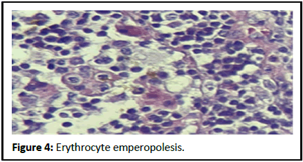
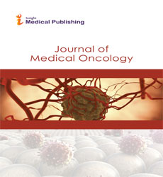
In the anatomopathological report it’s referred to a cervical adenomegaly, the two samples sent to analysis present a smooth external surface, of bright light brown color. At the cut, consistency is soft. The surface of the mayor node cut is made of smaller nodes that form a bigger node. The surface of the cut of the minor node is made of large and homogenous lobes of light brown color. Under the microscope we can observe a presence of emperipolesis of erythrocytes and some hemosiderophages (Figures 3 and 4).
Discussion
Rosai-Dorfman is an uncommon entity with scarce cases reported from its description, characterized by a bening proliferation of histiocytic cell located in the ganglionic sinuses or lymph vessels [2]. It presents itself in Caucasian origin patients and afro descendants, with the most affected age group being between the first and second decade of life [3]. The progress of the disease is chronic and tends to have alternating exacerbations with spontaneous remissions [4].
It has three ways of presenting itself: Nodal or classical, extra-nodal and cutaneous, with the first being the most frequent one. In the case of the nodal presentation, the clinical picture can go along with symptoms like fever and leukocytosis [5] and in some cases we can observe weight loss and nocturnal diaphoresis, although in most cases, the general state of the patient is kept in good shape. According to Cai, Shi and Bai [6] approximately 43% of the cases are of extra-nodal presentation in at least one location, mainly in head and neck, while only 23% have an exclusively extra-nodal presentation; the most common extra-nodal sites found are the respiratory tract, visceral organs, genitourinary system, skin, bone, SNC and soft tissue [6]. Manifestations like normocytic normochromic anemia have been observed in approximately 67% of cases, as has the presence of hypergammaglobulinemia, thrombocytopenia, eosinophilia and inflammatory syndrome. The neurological manifestations are more present in greater frequency on elderly patients, in an intercranial form, this type of manifestation could look clinically and radiographically like a meningioma [7] the lesions on bone usually are osteolytic affecting mainly long bones, vertebrae and sacrus [3].
Histopathologically we can observe an increase in size of the lymph nodes and dilation of the medullary and capsular sinuses, its characteristic the presence of emperipolesis in the cell of the cells in RDD [5,8]. The characteristics of the nodal presentation can be confused with those of the extra-nodal presentation; however, the latter usually have prominent lymphoid follicles with germinal centers, sclerosis, few histocytes and fibrosis, the emperipolesis is usually mild.
Its etiopathology is unknown, however, many theories allow us to get to plausible explanations, but until now, none have been conclusive. According to numerous studies, one of the possible mechanisms involved includes the participation of the immune system, specifically, an alteration in the correct regulation of an immunological response to various infectious agents, having as the most frequent example that of Eipsen-Barr virus [9]. It has also been suggested the participation of Klebsiella, Brucella or Cytomegalovirus as causing agents, the genotype 6 herpes virus was demonstrated in a biopsy sample via in situ hybridization [10].
Another aspect to consider is the presence and action of monocytes and cytokines. In Cai’s investigation [6] the expression of CD11c has been found in diverse macrophages, especially those with an inflammatory environment, this protein shares its expression with the histiocytes characteristic of RDD and the infiltration of cells that are present in lesions of such. Adding to this, the presence of Ly-6C was also found in rodents, which would indicate the capacity of monocytes to migrate to lymph nodes and induce the expression of CD8+. Therefore, the evidence of said study allow to conclude that, in presence of an adequate stimulus, the lymphocytes can act as antigen presenting cell and move to lymph nodes to make part of the infiltration characteristic of RDD.
Recently the mutation in kinases was described, same that could explain a clone nature, even though said possibility had been discarded earlier, it can be observed in the nodal presentations as well as in the extra-nodal presentation, only excluding the cutaneous presentation. These mutations include KRAS, MAP2K1, ARAF and NRAF, being found most frequently in MAP2K1 and KRAS, while being understood as a possible indication of the clone nature in a certain number of cases [11].
The differential diagnosis of the pathology is of uttermost importance, the sinus histiocytosis with massive lymphadenopathy shares histological characteristics with other types of systemic histiocytosis (Histiocytosis of Langerhans cells, Erdheim-Chester disease), for example, the presence of inflammatory cells, histiocyte infiltration on tissue that presents itself as marker of Langerhans cells as well as without them and in many cases, affecting various organs [3]. In order to differentiate RDD from the rest of histiocytosis, we can recur to immunohistochemistry; the histocytes that present themselves in Rosai-Dorfman Disease have a positive reaction to the S-100 protein but react negatively to CD1a and HLA-DR [6]. The presence of S-100+ but CD1a- histocytes with evidence of emperipolesis can work as a definitive diagnostic to identify RDD. Within the differential diagnosis there the inclusion of hemophagocytic syndrome, tuberculosis and lymphoma [10].
In the laboratory, patients with RDD have presented an increased rate of erythrocytic sedimentation, elevated IgC and positive Antinuclear Antibody (ANA). As it was mentioned before, there is observation of normochromic normocytic anemia, leukocytosis with neutrophilia and less frequently hemolytic anemia and eosinophilia [8]. The most common characteristic found in imaging is lymphadenopathy, same that can be disseminated, be isolated or be combined with extra-nodal characteristics [12]. In any case, the definitive diagnosis must always be based in a complete clinical history, a correct examination and be multidisciplinary, that is, include a complete analysis and exhaustive clinical, histopathological, laboratory and imaging evidence to establish and define the presence of RDD.
Rosai-Dorfman is a pathology of chronical and unpredictable evolution, there is no stablished ideal treatment, observation on asymptomatic or mild symptomatic patients is advised, as long as there are no lesions in vital organs and they are not massive [13]. The case election in case of symptomatic patients whose vital functions are endangered by the disease progress is surgery, patients with unresectable multifocal extra-nodal disease would require systemic treatment, which includes immunomodulating therapy, radiotherapy, chemotherapy and corticosteroids [11]. However, no treatment has been proven to be more effective than others.
The prognosis of the pathology depends on the amount of affected lymph nodes. Without the treatment the patients could present recurrence in approximately 70% of cases [8]. The objective of the treatment is to be the least invasive possible to control symptomatology and preserve the patient’s life [14,15].
Conclusion
Destombes-Rosai disease is a histiocytosis that presents itself with a lymphadenopathy that is commonly bilateral, painless accompanied with fever, despite being a rare pathology, it is benign and tends to be self-limited. Its most common presentation is nodal, but it can be systemic as well. We presented a case report with both a nodal and systemic presentation where imaging reveals multiple adenopathy in all ganglionic levels with atypical characteristics. The purpose of this report is denote the presence of both presentations in a pediatric patient, considering Rosai-Dorfman disease as a differential diagnosis of importance while observing lymphadenopathies.
Consent
Written informed consent for publication of their clinical details and/or clinical images was obtained from the parent(s)/patient. All the information provided has been completely attached to national and international ethics standards, reviewed by the hospital and informed to the parent(s)/patient. There is permission to reproduce the data from other sources, with parent(s)/patient approval as well.
Funding
There are/were no funding and/or conflicts of interests/competing interests in any of the provided information.
Data is available at the hospital Para el Niño Poblano, should it be properly requested with parent(s)/patient consent and a well organised statement with the reasons for the acquisition.
All the obtained material such as blood samples, laboratory data, presented images, tissue samples and every documented evidence are declared in the informed consent provided by the hospital with the parent(s)/patient(s) signature/approval with the hospital´s ethics committee knowledge and approval as well.
Acknowledgement
Not applicable.
References
- Aubart FC, Haroche J, Emile JF, Charlotte F, Barete S, et al. (2018) Destombes-Rosai-Dorfman disease: Evolution of the concept, classification and management. J Intern Med 39:635-640
- Galicier L, Fieschi C, Meignin V, Clauvel JP, Oksenhendler E (2007) Rosai-Dorfman sinus histiocytosisRosai-Dorfman disease. Med press 36:1669-1675
- Papo M, Cohen-Aubart F, Trefond L, Bauvois A, Amoura Z, et al. (2019) Systemic histiocytosis (Langerhans cell histiocytosis, Erdheim–Chester disease, Destombes-Rosai-Dorfman disease): From oncogenic mutations to inflammatory disorders. Curr Oncol Rep 21:1-10
[Crossref] [Google Scholar] [PubMed]
- Shrirao N, Sethi A, Mukherjee B (2016) Management strategies in Rosai-Dorfman disease: To do or not to do. J Pediatr Hematol Oncol 38:e248-e250
[Crossref] [Google Scholar] [PubMed]
- Kaltman JM, Best SP, McClure SA (2011) Sinus histiocytosis with massive lymphadenopathy Rosai-Dorfman disease: A unique case presentation. Oral Surg Oral Med Oral Pathol Oral Radiol Endod 112:e124-e126
[Crossref] [Google Scholar] [PubMed]
- Cai Y, Shi Z, Bai Y (2017) Review of Rosai-Dorfman disease: New insights into the pathogenesis of this rare disorder. Acta Haematol 138:14-23
[Crossref] [Google Scholar] [PubMed]
- Tomio R, Katayama M, Takenaka N, Imanishi T (2012) Complications of surgical treatment of Rosai-Dorfman disease: A case report and review. Surg Neurol Int 3:1
[Crossref] [Google Scholar] [PubMed]
- Mi3kus A, Stefanowicz J, Kobierska-Gulida G, Adamkiewicz-Drozynska E (2018) Rosai-Dorfman disease as a rare cause of cervical lymphadenopathy-case report and literature review. Cent Eur J Immunol 43:341-345
[Crossref] [Google Scholar] [PubMed]
- Azari-Yaam A, Abdolsalehi MR, Vasei M, Safavi M, Mehdizadeh M (2021) Rosai-Dorfman disease: A rare clinicopathological presentation and review of the literature. Head Neck Pathol 15:352-360
[Crossref] [Google Scholar] [PubMed]
- Kumar B, Karki S, Paudyal P (2008) Diagnosis of sinus histiocytosis with massive lymphadenopathy (Rosai-Dorfman disease) by fine needle aspiration cytology. Diagn Cytopathol 36:691-695
[Crossref] [Google Scholar] [PubMed]
- Bruce-Brand C, Schneider JW, Schubert P (2020) Rosai-Dorfman disease: An overview. J Clin Pathol 73:697-705
[Crossref] [Google Scholar] [PubMed]
- Vaidya T, Mahajan A, Rane S (2020) Multimodality imaging manifestations of Rosai-Dorfman disease. Acta Radiol Open 9:2058460120946719
[Crossref] [Google Scholar] [PubMed]
- Pulsoni A, Anghel G, Falcucci P, Matera R, Pescarmona E, et al. (2002) Treatment of sinus histiocytosis with massive lymphadenopathy (Rosai-Dorfman disease): Report of a case and literature review. Am J Hematol 69:67-71
[Crossref] [Google Scholar] [PubMed]
- Kutlubay Z, Bairamov O, Sevim A, Demirkesen C, Mat MC (2014) Rosai-Dorfman disease: A case report with nodal and cutaneous involvement and review of the literature. Am J Dermatopathol 36:353-357
[Crossref] [Google Scholar] [PubMed]
- Parkash O, Yousaf MS, Fareed G (2019) Rosai-Dorfman's disease, an uncommon cause of common clinical presentation. J Pak Med Assoc 69:1213-1215
[Google Scholar] [PubMed]
Open Access Journals
- Aquaculture & Veterinary Science
- Chemistry & Chemical Sciences
- Clinical Sciences
- Engineering
- General Science
- Genetics & Molecular Biology
- Health Care & Nursing
- Immunology & Microbiology
- Materials Science
- Mathematics & Physics
- Medical Sciences
- Neurology & Psychiatry
- Oncology & Cancer Science
- Pharmaceutical Sciences


