ISSN : ISSN: 2576-1412
Journal of Applied Microbiology and Biochemistry
Report of a Case of Human Nasal Myiasis Caused By Second Instar Larvae of Oestrus Ovis in CHU Oran: Review of Literature
Pr Benmansour Zakaria1* and Raiah Abdelhalim2
1Department of Parasitology and Mycology, University of Es Senia, Algeria
2Medecin Resident in Parasitology and Mycology, CHU Oran, Algeria
- *Corresponding Author:
- Pr Benmansour zakaria
Department of Parasitology and Mycology, CHU Oran
University of Es Senia/Faculty of Medicine
Oran, Algeria
Tel: 00213770321576
E-mail: benzakarion31@yahoo.fr
Received Date: May 09, 2017; Accepted Date: May 19, 2017; Published Date: May 29, 2017
Citation: Zakaria PB, Abdelhalim R. Report of a Case of Human Nasal Myiasis Caused By Second Instar Larvae of Oestrus Ovis in CHU Oran: Review of Literature. J Appl Microbiol Biochem. Vol. 1 No. 3:9
Abstract
Myiasis is the invasion of human and animal organs and tissues by dipteran larvae. This is a case report of a human nasal myiasis caused by the second instar larvae of the sheep bot fly, Oestrusovis. The cases of human myiasis at Oestrusovis are accidental and rare, mainly affecting the eyes. In the literature, there are only sporadic reports of nasal myiasis.
We present a new case of nasal myiasis caused by the second instar larvae of Oestrusovis in a 29-year-old male patient, originally from OuedRhiou in the west of Algeria, whois a medical doctor suffering from signs of rhinosinusitis.
Keywords
Oestrusovis; Human myiasis; Nasal myiasis
Introduction
Myiasis is defined as the invasion of human and animal organs and tissues by dipteran larvae [1-4] which optionally or necessarily feed on living or necrotic tissuesfor at least a period of time [5]. This is an essentially veterinary pathology. The human being is infected only accidentally [6-8]. Nasal myiasis is much more prevalent in tropical countries. The localizations in humans are varied: Cutaneous and subcutaneous, gastrointestinal, genitourinaryor cavities of the face that can cause damage to the nasal cavities, ear canal or eyes [8] Clinically, human myiasis may be benign and asymptomatic, or on the contrary may lead to violent disturbances or even death [9,10]. Among these types of myiasis, the nasal form is relatively rare [11,12]. We describe a nasal myiasis due to Oestrus ovis in a 29 year old patient from OuedRhiou, a doctor by profession with a literature review.
Case Presentation
A 29-year-old male patient from OuedRhiou who lives in Oran, Algeria, Without special medical history, Showing signs of left Rhinosinusitis with nasal discharge abundant two weeks after a short stay in his native village (OuedRhiou) 140 km east of Oran. Five days after the onset of this symptom and following a sneeze, two larvae are expelled from his nose, bringing him to consult the Department of Medical Parasitology and Mycology of CHU d'Oran. An endoscopy carried out at the ENT department showed a slight inflammation in the nasopharynx and did not visualize any other residual larvae, an antibiotic treatment is then instituted in order to avoid superinfection.
Ten days later, the patient brought us a larva expelled from his nose. Fixed to 70% alcohol, and then heated to 90°C with 5% glycerine in our service for identification [13]. The patient's biological assessments were unusual, the frontal X-ray showed a small left maxillary sinus filling, supplemented by a CT scan of the sinuses, which objectified an aspect of a thickening of the left maxillary sinus mucosa, evoking chronic sinusitis.
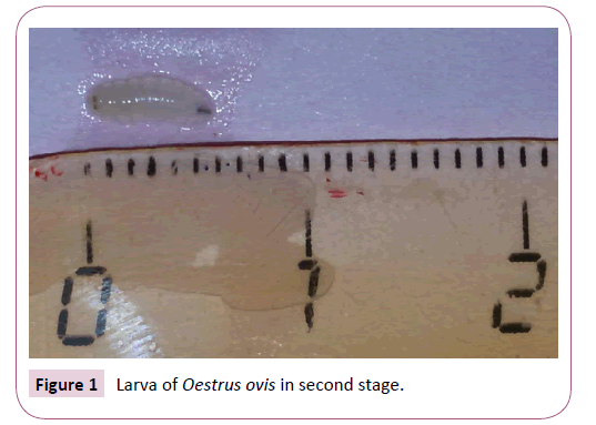
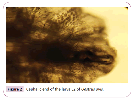
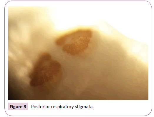
The larva was identified macroscopically and microscopically according to the Zumpt criteria [1]. This was the second stage L2 larva of Oestrus ovis, measuring 6 mm long. This larva is of semicylindrical shape (Figure 1). The anterior end or pseudocephalon has two visible buccal hooks (Figure 2). The last metamerus carries the posterior sub-circular stigmata with a central button, Pierced with numerous pores [1-14] (Figure 3).
Discussion
The human being is not a usual host of the Oestrus ovis fly or sheep nasal bot fly, but rare cases of larval infestations of these diptera have been described. The eye is the most affected site, but larvae have been unusually found in human nasal cavities [4]. Even rarer are the cases of nasal myiasis with development of larvae up to the second and third stages.
We carried out a bibliographic search on Pub Med describing and listing all cases of human nasal myiasis due to Oestrus ovis in the literature, in total, eighteen are the publications that report in detail twenty-one cases of human nasal myiasis due to Oestrus ovis, presented in Table 1 among seventy publications of cases of human nasal myiasis due to different species of diptera larvae, Oestrusovis included.
Table 1 Cases of human nasal myiasis due to Oestrus ovis reported in the world, since 1958
| Years | Countries | Number of Cases | Notes | References |
|---|---|---|---|---|
| 1958 | Algeria | 1 case | [19] | |
| 1977 | Turkey | 1 case | [20] | |
| 1978 | India | 1 case | [21] | |
| 1990 | Spain | 2 cases | [11] | |
| 1997 | United Kingdom | 1 case | [22] | |
| 1997 | Egypt | 2 cases | [23] | |
| 1999 | New Zeland | 2 cases | Associatedwithophthalmomyiasis | [24] |
| 2001 | France | 1 case | [4] | |
| 2007 | China | 1 case | Location at the maxillary sinus | [25] |
| 2007 | CanaryIslands | [26] | ||
| 2008 | Turkey | 1 case | [27] | |
| 2010 | United Kingdom | 1 case | Associatedwithophthalmomyiasis | [28] |
| 2011 | Morocco | 1 case | [29] | |
| 2011 | Sweden | 1 case | [30] | |
| 2011 | Israel | 1 case | [31] | |
| 2012 | Neterlands | 1 case | Location at the maxillary sinus | [32] |
| 2015 | China | 1 case | [33] | |
| 2015 | CanaryIslands | 1 case | Associatedwithophthalmomyiasis | [34] |
It is in Algeria, since 1904, that the brothers SERGENT have for the first time demonstrated the role of fly larvae Oestrus ovis in human pathology with agent responsible for ophthalmomyiasis [15-17] a country where the human myiasis had been known since the beginning of time by the inhabitants who called them thimni, which is that of the sheepbot fly in the Berber dialects [15,17]. In 1952, Edmond SERGENT published a survey on the geographical distribution of this human oculo-nasal myiasis due to Oestrisovis in the world and proposed the use of thimni names for myiasis [18]. The cases of human myiasis described in the literature in Algeria are shown in Table 2 the actual number of cases is certainly higher than the published cases, which makes it difficult to appreciate the actual incidence of myiasis in Algeria.
Table 2 Cases of human myiasis reported in Algeria, since 1929
| Years | Type of Myiasis | Species | Number of Cases | References |
|---|---|---|---|---|
| 1929-1952 | Ophthalmomyiasis | Oestrusovis | 50 cases | [18] |
| 1958 | Oculo-nasal myiasis | Oestrusovis | 1 case | [19] |
| 1991 | Head skin myiasis | Lucilia | 2 cases | [35] |
| 1999 | Urogenitalmyiasis | Faniacanicularis | 1 case | [36] |
| 1997 | Auricularmyiasis | Chrysomabezziana | 1 case | [17] |
Oestrus ovisis a yellowish-gray, cosmopolitan, non-pungent dipteran, 10-12 mm long. The adult has a short life devoted solely to breeding during which he does not feed. Larvae are obligatory parasites of the nasal fossae and frontal sinuses of sheep and goats [6-8]. Oestrus ovis' female viviparous fly, often in flight, deposits its larvae at the nostrils of the mammal [18-39]. First instar larvae deposited in the autumn (September to October) move up the animal's nasal fossae to reach the frontal sinuses where they mature to reach the third larval stage in two to twelve months and will be released into the nasal mucus the following spring [4]. They fall or are eliminated during sneezing on the soil, where they are transformed into a pupa, from which an adult insect is immersed four to six weeks later and the cycle begins again [4,6,8,15,37,39,40,41]. The animals thus infected show a summer rhinitis followed by a winter sinusitis [4].
In sheep, the pathogenic action results from two phenomena, namely a minor mechanical effect and allergic hyper-sensitization phenomena [4,14,29,38,42,43]. In humans, the traumatic and mechanical pathogenic action is preponderant. Clinical signs are due to the presence of larvae with buccal hooks and numerous very strong spines which ensure both their fixation and their displacement [24,26,38]. The short duration of parasite infestation in humans would prevent allergic expression of larvae [4,24,26,29,38].
The infestation of man is rare, it generally interests the eye [4]. The contamination is made from larvae L1 stages which are deposited by the female fly in full flight at the level of the conjunctival sac [6,27,37]. The human feels a brutal shock on his eye followed by inflammation, some hour later. The evolution is favourable after the extraction of the larvae [4,37,40-47]. Exceptionally, L1 larvae may settle at the external nasal orifices causing an itching sensation, boots of sneezing accompanied by an abundant nasal discharge, seeing pains in relation to the sinuses [4,18]. Note also the possibility of deposition of the larvae on the lips responsible for oropharyngealmyiasis. In the cases reported, the duration of the pathology generally does not exceed fifteen days and the symptoms disappear gradually after the extraction of the larvae [4].
Treatment consists of larval excision, antibiotic treatment may be combined to avoid superinfection [4,24,37,41,46], some authors recommend the systemic use of ivermectin which is an anthelmintic treatment [48].
The development of rhinomyiasis is usually benign after treatment. However complications have been described, as in the case of a patient with a nasal myiasis complicated by apneumocephalus [10].
Conclusion
Nasal myiasis is relatively rare in humans and can have consequences ranging from simple rhinitis and psychological stress to lifethreatening complications of the patient [9]. It is necessary to know to evoke them in front the sudden appearance of the signs of rhinosinusitis with sensation of a foreign body moving in the nasal cavities, in order to be able to treat them early and to avoid the complications that can occur.
References
- Zumpt F (1965)Myiasis in man and animals in the old world: a textbook for physicians, veterinarians and zoologists. Butterworth Co Ed, London. p. 257.
- Lapierre J, Pette M (1954) À propos d'un cas de myiase oculaire dû à Oestrusovis observé dans la région parisienne. Bulletin de la Société de Pathologie Exotique 47: 561-563.
- Steiner C, Deduit Y (1959) Myiase oculaire. Bulletin des Sociétés d’Ophtalmologie Françaises 12: 924-927.
- Delhaes L, Bourel B, Pinatel F, Cailliez JC, Gosset D, et al. (2001) Myiasenasalehumaine à Oestrusovis. Parasite 8: 289-296.
- Service MW (1981) Medical entomology for students.Cambridge University Press, Cambridge.
- Ducourneau D (1981) Les myiases oculaires. Med Trop 41:511-514.
- Rodhain F, Perez C (1985) Les diptères myiasigènes. In Précis d’entomologie médicale et vétérinaire. Maloine, Paris. pp: 249-265.
- Anane S, Ben Hssine L (2010) La myiase conjonctivale humaine àOestrusovisdans le sud tunisien. Bull SocPatholExot103:299-304.
- Wood TR, Slight JR (1970)Bilateral orbital myiasis: Report of a case. Arch Ophthalmol84:6923.
- Kuruvilla G, Albert RRA, Job A, Ranjith VT, Selvakumar P (2006)Pneumocephalus: A rare complication of nasal myiasis. Am J Otolaryngol27: 133-135.
- Quesada P, Navarrete ML, Maeso J (1990) Nasal myiasis due to Oestrusovislarvae. Eur Arch Otorhinolaryngol247: 131-132.
- Babamahmoudi F, Rafinejhad J, Enayati A (2012) Nasal myiasis due to Luciliasericata (Meigen, 1826) from Iran: A case report. IRAN Tropical Biomedicine 29: 175-179.
- Garcia LS, Bruckner DA (1988) Diagnostic medical parasitology. Elsevier Science Publishing, NewYork. p. 431.
- Gianneto S, Santoro V, Pampiglione S (1999) Scanning electron microscopy ofOestrusovislarvae (Diptera: Oestridae): Skin armour and posterior spiracles. Parasite 6: 73-77.
- Sergent ED, Sergent ET (1907) Myiase humaine d’Algérie causée par Oestrusovis. Ann Inst Pasteur 21: 392-399.
- Sergent ED, SergentT ET(1913) La «Tamné». Myiase humaine des montagnes sahariennes touarègues, identique à la «Thimni» des Kabyles, due à Oestrusovis. Bull Soc PathExot 6: 487-488.
- Abed-Benamara M, Achir I, Rodhain F, Perez-Eid C (1997) Premier cas algerien d’otomyiase humaine à Chrysomyabezziana.Bull Soc PatholExot 90:172-175.
- Sergent E, La Thimni(1952) Myiase oculo-nasale de l’homme causée par l’œstre du mouton. Arch InstPasteur Alger 15: 319-322.
- Favier G (1958) A case of oculo-nasal myiasis caused by Oestrusovis and seen at Saoura (southern Oran).Arch Inst Pasteur Alger 36:182-183.
- Yalçinkaya F (1977)A case of nasomyiasis caused by Oestrusovis larva.Turk HijTecrBiyolDerg 37:262-265.
- Agrawal RD, Ahluwalia SS, Bhatia BB (1978) Occurrence of Oestrusovis larvae in nasal cavity of a woman. J Indian Med Assoc 70: 263.
- Yeruham I, Malnick S, Bass D, Rosen S (1997) An apparently pharyngeal myiasis in a patient caused by Oestrusovis (Oestridae: Diptera). Acta Trop 68: 361-363.
- Lucientes J, Clavel A, Ferrer-Dufol M, Valles H, Peribanez MA, et al. (1997) Short report: One case of nasal human myiasiscaused by third stage instar larvae of Oestrusovis. Am J Trop Med Hyg56: 608-609.
- Fekry AA, el Serougi AO, Ayoub SA(1997) Oestrusovis (sheep nasal fly) infesting the eyes and the nose of a camel keeper family. J Egypt SocParasitol 27:493-496.
- Macdonald PJ, Chan C, Dickson J, Jean-Louis F, Heath A (1999)Ophtalmomyiasis and nasal myiasis in New Zealand: a case series. NZ Med J 112: 445-447.
- Zhang XC, Liu XD, Lu ZM (2007)Myiasis of maxillary sinus and nasal cavity by the larvae of Oestrus ovis.ZhongguoJi Sheng Chong XueYuJi Sheng Chong Bing ZaZhi25:1.
- Hemmersbach-Miller M, Sánchez-Andrade R, Domínguez-Coello A, Meilud AH, Paz-Silva A, et al. (2007) HumanOestrussp. Infection, CanaryIslands. Emerg Infect Dis 13:950-952.
- Eyigör H, Dost T, Dayanir V, Başak S, Eren H (2008) A case of naso-ophthalmic myiasis.Kulak BurunBogazIhtisDerg18:371-373.
- Smillie I, Gubbi PK, Cocks HC (2010) Nasal and ophthalmomyiasis: case report. J LaryngolOtol 124:934-935.
- Tligui H, Oudaina W, Khairane I, Boughaidi A, Lamalmi F, et al. (2011)Rhino-myiase humaine à Oestrusovis: Une observation au Maroc. Med Trop (Mars) 71:83-84.
- Einer H, Ellegard E (2011) Nasal myiasis by Oestrus ovis second stage larva in an immunocompetent man: case report and literature review. J LaryngolOtol125:745-746.
- Mumcuoglu KY, Eliashar R (2011) Nasal myiasis due to Oestrus ovis larvae in Israel. Isr Med Assoc J 13:379-380.
- Hummelen R, Zeegers T, den Hollander J, Tabink I, Ten Koppel P (2012)Eenongewoneoorzaak van sinusitis.Ned TijdschrGeneeskd 156:A5373.
- Su H, Liu L, Zhao Y (2015) One case of human nasal myiasis. Lin Chung Er Bi Yan HouTou Jing WaiKeZaZhi29: 1138-1139.
- SanteFernández L, Hernández-Porto M, Tinguaro V, LecuonaFernández M (2015)Ophthalmomyiasis and nasal myiasis baaay Oestrus ovis in a patient from the Canary Islands with uncommon epidemiological characteristics.EnfermInfeccMicrobiol Clin 24: 213-375.
- BoudgheneSO, MeradBA (1991) Myiase de plaies du cuir chevelu chez deux enfants de Tlemcen (Algérie). Bull Soc PatholExot 84:283-285.
- Perez-Eid C, Mouffok N (1999) Myiase urinaire humaine a larves de Fanniacanicularis (Diptera, muscidae) en Algérie. Presse Med 28:580-581.
- Zayani A, Chaabouni M, Gouiaa R, HamidaFBH,Fki J (1989) Myiases conjonctivales. À propos de 23 cas dans le Sahel tunisien. ArchsInst Pasteur Tunis 66: 289-292.
- Dorchies P (1997) Physiopathologie comparee de la myiase aOestrusovis (Linné 1761) chez l’homme et chez les animaux. Bull AcadNatl Med 181: 673-84.
- Jacquiet P (1997) Oestrus ovis.Le Point Veterinaire28: 31-32.
- ViallefontH,Boudet C, Jarry D (1961)Ophtalmomyiase par Oestrusovis(Etude parasitologique).Bulletin des Sociétés d'Ophtalmologie Françaises1: 256-263.
- Mariotti JM, Vacheret G (1992) Les myiases conjonctivales, une pathologie fréquente en Corse. J Fr Ophtalmol 15: 679-682.
- Nglyen VK, Jacquet P, Duranton C, Bergeaud JP, Prevot F, et al. (1999) Reactions cellulaires des muqueuses nasales et sinusales des chèvres et des moutons à l'infestation naturelle par Oestrusovis Linné175 8 (Diptera: Oestridés). Parasite 2: 141-149.
- Khan ZA, Al-Jama AA, Rahi AHS, Ahmed M, Madan I (1993) Myiasis: Report of two cases. Annals of Saudi Med13: 464-466.
- Dorchies P, Larrouy G, Deconinck P, Chantal J (1995) L’ophtalmomyiase externe humaine : Revue bibliographique a propos de cas en République de Djibouti. Bull Soc PatholExot 88: 86-89.
- Cohen H, Rozenman Y, Ronen S (1981) Trois cas de myiases conjonctivales dues à Oestrusovis. J Fr Ophtalmol 4: 583-585.
- Delord JJ (1976) Une parasitose de chez nous : l’euliase de la conjonctive oculaire. MediterraneeMedic 106: 31-32.
- Shinohara EH,Martini MZ, de Oliveira NetoHG,Takahashi A (2004) Oral myiasis treated with ivermectin: Case Report. Brazilian Dental J 15: 79-81.
Open Access Journals
- Aquaculture & Veterinary Science
- Chemistry & Chemical Sciences
- Clinical Sciences
- Engineering
- General Science
- Genetics & Molecular Biology
- Health Care & Nursing
- Immunology & Microbiology
- Materials Science
- Mathematics & Physics
- Medical Sciences
- Neurology & Psychiatry
- Oncology & Cancer Science
- Pharmaceutical Sciences


