Renal Leiomyosarcoma: A Rare Neoplasm of Kidney
Joydeep Singh Vasant*, Ajay Yadav, Neha Sethi, Manish Vijayvargiya, Tarun Jain, Hemant Malhotra
Department of Science, Maharishi Markandeshwar University, Haryana, India
- *Corresponding Author:
- Joydeep Singh Vasant
Department of Science,
Maharishi Markandeshwar University,
Haryana,
India,
Tel: 8745004612;
Email: jsvasant@gmail.com
Received date: October 18, 2022, Manuscript No. IPMO-22-14823; Editor assigned date: October 21, 2022, PreQC No. IPMO-22-14823 (PQ); Reviewed date: November 07, 2022, QC No. IPMO-22-14823; Revised date: December 27, 2022, Manuscript No. IPMO-22-14823 (R); Published date: January 02, 2023, DOI: 10.36648/IPMO.6.1.001
Citation: Vasant JS, Yadav A, Sethi N, Vijayvargiya M, Jain T, et al. (2023) Renal Leiomyosarcoma: A Rare Neoplasm of Kidney. J Med Oncol Vol:6 No: 1
Abstract
Leiomyosarcoma of the kidney is a rare entity, accounting for only less than 1% of renal malignancies. Only few cases have been reported in literature. Radiologically it is difficult to distinguish them from renal cell carcinoma. Accurate diagnosis is mandatory for proper management. Histopathological examination and Immune Histo Chemistry (IHC) are required to establish a diagnosis. We are presenting a case of renal leiomyosarcoma in a 45 years old male.
Keywords
Immune Histo Chemistry (IHC); Leiomyosarcoma; Lymphnodes; Chemotherapy; Hematuria
Introduction
Sarcomas represent less than a fraction of a percent of overall malignancies. They are a part of a broad group of neoplasms stemming from mesenchymal origin with greater than 70 differentiating histological classifications. Leiomyosarcoma occurs secondary to malignant transformation of smooth muscle cells in our body. Leiomyosarcoma of the kidney is an extremely rare tumor, accounting for only 0.12% of renal malignancies. The exact etiology is unknown, however like many other cancers, genetic and environmental factors can likely be attributed. It is known to preferentially affect females in their 60’s. The symptoms and signs of this tumor are non-specific as patients usually complain of flank pain or abdominal pain and hematuria [1]. Aggressive course and the potential role of therapy in a metastatic setting mandate our understanding of this pathology. The current treatment of choice of leiomyosarcoma is radical nephrectomy. Making an accurate preoperative diagnosis may be beneficial as neoadjuvant chemotherapy can be utilized to treat potential micrometastasis. Moreover, adjuvant chemotherapy and radiotherapy maybe used in the care of patients with renal leiomyosarcoma. After surgery patients need adjuvant therapy to reduce the risk of recurrence. Metastatic disease is associated with poor prognosis. We present a case of inoperable metastatic leiomyosarcoma in a 45 years old male patient.
Case Presentation
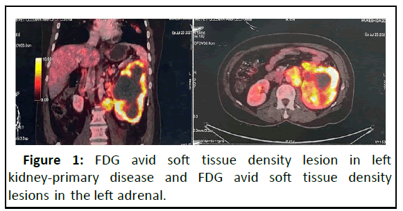
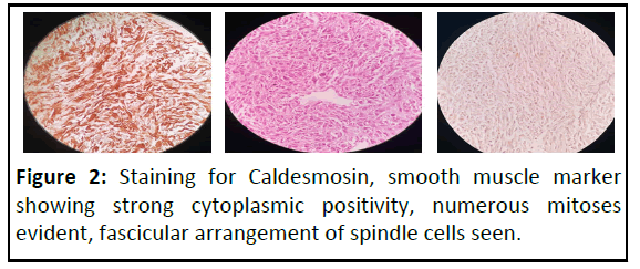
A 45 years old male presented with a 3 months history of abdominal pain and cough. Ultrasound abdomen and pelvis revealed a solid well defined hypoechoic mass arising from the left kidney and contrast enhancing CT scan abdomen and pelvis revealed an enhancing mass in the left kidney, hyperattenuating in comparison to the renal parenchyma [2]. The core biopsy of the left renal mass revealed high grade sarcoma, IHC-Vimentin, SMA, Desmin, H-caldesmon: Positive and pancytokeratin, CD-34, S-100, TLE: Negative, the final impression revealed leiomyosarcoma. A whole body PET-CT scan was done for metastatic workup which revealed FDG avid soft tissue density mass in the left kidney of size 10.5 cm × 11.4 cm × 16.7 cm with metastasis to paraaortic and preaortic region, bilateral supraclavicular, mediastinal, abdominal lymphnodes, lung nodules and skeletal lesions in L4 and D10 vertebra and left adrenal lesion. The case was discussed in multidisciplinary tumor board and planned for palliative chemotherapy with bisphosphonate for bony metastasis. The patient was given 3 cycles of chemotherapy with Ifosfamide and Doxorubicin, along with Zolendronic acid. The interim PET CT scan after 3 cycles of chemotherapy revealed partial response. Planned to give 3 more cycles of chemotherapy (Figures 1 and 2).
Figure 2: Staining for Caldesmosin, smooth muscle marker showing strong cytoplasmic positivity, numerous mitoses evident, fascicular arrangement of spindle cells seen.
Discussion
Leiomyosarcomas are malignant neoplasms of smooth muscle origin. Apart from the uterus, soft tissue leiomyosarcomas commonly occur in the retroperitoneum and usually involves the lower extremity but they can occur in the head and neck region also. Primary leiomyosarcomas are rare in the kidneys and constitute 0.12% of all malignant renal tumors but they are the commonest renal sarcomas [3].
Primary leiomyosarcoma of the kidney has preponderance in women and is more frequent in fifth decade but can be found in almost any age group in the later period of life and is more common in the right as compared to the left kidney. The life expectancy is lower with the sarcomas of the kidney as compared to sarcomas of the other parts of the urinary tract [4]. The histogenesis of leiomyosarcomas is believed to be from the renal capsule or the smooth muscle fibres in the renal pelvis or from the renal vessels [5]. The most common clinical symptoms are pain, palpable mass and hematuria, all of which are indicators of extensive local disease [6,7].
Grossly renal leiomyosarcomas may appear as evident trabaculations and who ling patterns, microscopically the tumor shows alternating fascicles of spindle shaped cells with marked nuclear atypia and prominent mitotic figures [8-11]. They are distinguished from renal leiomyomas by cellular pleomorphism, increased mitotic rate and the presence of cellular necrosis. On immunohistochemistry they show a positive reaction for smooth muscle markers like SMA with negative cytokine markers [12-14]. Immunohistochemical examination is also used to differentiate leiomyosarcoma from sarcoid renal cell carcinoma when distinction can be difficult based solely on histological comparisons [15].
Conclusion
Resection is the standard treatment for these aggressive lesions, followed by either chemotherapy or radiotherapy. In general there is no survival advantage with adjuvant/ neoadjuvant chemotherapy/radiotherapy in leiomyosarcoma however Raut. reported that adjuvant chemotherapy could be used on partially resected tumors and Naguma et al reported incomplete response in metastatic renal leiomyosarcoma with systemic chemotherapy.
Renal leiomyosarcomas have a very poor prognosis because of aggressive behavior owing to rapid growth rate and high potential for metastasis.
References
- Niceta P, Lavengood RW Jr, Fernandes M, Tozzo PJ (1974) Leiomyosarcoma of kidney: Review of the literature. Urology 3:270-277
[Crossref] [Google Scholar] [PubMed]
- Angel CA, Gant LL, Parham DM, Rao BN, Douglass EC, et al. (1992) Leiomyosarcomas in children: Clinical and pathologic characteristics. Pediatr Surg International 7:116-120
- Dhawan S, Chopra P, Dhawan S (2012) Primary kidney leiomyosarcoma. A diagnostic challenge. Urol Ann 4:48-50
[Crossref] [Google Scholar] [PubMed]
- Beardo P, Jose Ledo M, Jose Luise RC (2013) Renal leiomyosarcoma. Rare Tumours 5:42.
- Lee G, Lee SY, Seo S, Jeon S, Lee H, et al. (2011) Prognostic factors and clinical outcomes of urological soft tissue sarcomas. Korean J Urol 52:669-673
[Crossref] [Google Scholar] [PubMed]
- Kavantzas N, Pavlopoulos PM, Karaitianos I, Agapitos E (1999) Renal leiomyosarcoma: Report of three cases and review of the literature. Arch Ital Urol Androl 71:307-311
[Google Scholar] [PubMed]
- Lacquaniti S, Destito A, Candidi MO, Petrone D, Weir JM, et al. (1998) Two atypical cases of renal leiomyosarcoma: Clinical picture, diagnosis and therapy. Arch Ital Urol Androl 70:199-201
[Google Scholar] [PubMed]
- Deyrup AT, Montgomery E, Fisher C (2004) Leiomyosarcoma of the kidney: A clinicopathologic study. Am J Surg Pathol 28:178–182
[Crossref] [Google Scholar] [PubMed]
- Pong YH, Tsai VFS, Wang SM (2010) Primary leiomyosarcoma of kidney. Formos J Surg 45:124-126
- Lehnhardt M, Muehlberger T, Kuhnen C, Brett D, Steinau HU, et al. (2005) Feasibility of chemosensitivity testing in soft tissue sarcomas. World J Surg Oncol 3:20
[Crossref] [Google Scholar] [PubMed]
- Moureau-Zabotto L, Thomas L, Bui BN, Chevreau C, Stockle E, et al. (2004) Management of soft tissue sarcomas in first isolated local recurrence: A retrospective study of 83 cases. Radiother Oncol 8:279-287
[Crossref] [Google Scholar] [PubMed]
- Youssef E, Fontanesi J, Mott M, Kraut M, Lucas D, et al. (2002) Long-term outcome of combined modality therapy in retroperitoneal and deep-trunk soft tissue sarcoma: Analysis of prognostic factors. Int J Radiat Oncol Biol Phys 54:514-519
[Crossref] [Google Scholar] [PubMed]
- Raut CP, Pisters PW (2006) Retroperitoneal sarcomas: Combined modality treatment apporoaches. J Surg Oncol 94:81-87
[Crossref] [Google Scholar] [PubMed]
- Nagumo Y, Kimura T, Ichioka D, Uchida M, Oikawa M, et al. (2013) Long-term survival with gemcitabine and docetaxel for renal leiomyosarcoma: A case report. Hinyokika Kiyo 59:497-510
[Google Scholar] [PubMed]
- Dominici A, Mondaini N, Nesi G, Travaglini F, Di Cello V, et al. (2000) Cystic leiomyosarcoma of the kidney: An unusual clinical presentation. Urol Int 65:229-231
[Crossref] [Google Scholar] [PubMed]
Open Access Journals
- Aquaculture & Veterinary Science
- Chemistry & Chemical Sciences
- Clinical Sciences
- Engineering
- General Science
- Genetics & Molecular Biology
- Health Care & Nursing
- Immunology & Microbiology
- Materials Science
- Mathematics & Physics
- Medical Sciences
- Neurology & Psychiatry
- Oncology & Cancer Science
- Pharmaceutical Sciences


