Incidental Finding of Pulmonary Azygos Lobe and Imaging Findings: A Case Report
Klenam Dzefi-Tettey*
Department of Radiology, Korle-Bu Teaching Hospital, Guggisberg Avenue, Accra, Ghana
- *Corresponding Author:
- Dzefi-Tettey K
Department of Radiology, Korle-Bu Teaching Hospital, Guggisberg Avenue, Accra, Ghana
Tel: +233244234399
E-mail: k.dzefitettey@kbth.gov.gh
Received date: September 09, 2019; Accepted date: September 19, 2019; Published date: September 30, 2019
Citation: Dzefi-Tettey K (2019) Incidental Finding of Pulmonary Azygos Lobe and Imaging Findings: A Case Report. Journal of Clinical Radiology and Case Report Vol.3 No.1:7.
Copyright: © 2019, Dzefi-Tettey K. This is an open-access article distributed under the terms of the Creative Commons Attribution License, which permits unrestricted use, distribution, and reproduction in any medium, provided the original author and source are credited.
Abstract
A 42-year-old male presented with dyspnoea post-surgery for a benign brain tumour and was referred to the radiology department for pulmonary CT angiography on account of suspected pulmonary embolism.
The CT pulmonary angiogram showed filling defects in the RT and LT main pulmonary arteries, their segmental and subsegmental arteries confirming pulmonary embolism. Incidentally visualized is a horizontally uprooted azygos vein making a profound pleural gap into the apical fragment of the correct upper flap making an Azygos projection. An Azygos projection is a typical anatomic variation of the correct upper projection because of invagination of the azygos vein and pleura during embryological advancement in the hatchling. It's anything but a genuine extra projection as it doesn't have its possess bronchus. Azygos lobe can mimic various conditions such as bulla or abscess, pulmonary nodule and lung mass. Hence, radiologists, thoracic surgeons, and respiratory care physicians should be aware of this anatomical variant and the clinical implications of azygos lobe as this can compromise the success of thoracoscopic procedures for sympathectomy or thoracic surgery.
Keywords
Azygos lobe; Azygos fissure; Lung; Pulmonary Computerized Tomography (CT); Angiography
Introduction
An azygos lobe is an unusual but significant anatomical variant of the right lung anatomy [1,2]. It was first recognized at autopsy by Heinrich Wrisberg in 1877 and was first described on the chest radiograph in 1923[1,3]. Azygos lobe is formed when a displaced right posterior cardinal vein penetrates into the zenith of the lung rather than typical relocation over it during embryogenesis [4]. Its worldwide incidence ranges between 0.2%-1.2% with approximately 0.4% on chest radiograph and 1% on anatomical dissection [5,6]. Azygos lobe is more common in males than females and there is a predilection for family inheritance [7-9]. Clinically, azygos lobe can mimic various conditions such as bulla, abscess, pulmonary nodule, lung mass and even a case of primary adenocarcinoma arising from an azygos projection has been announced. They are usually found incidentally during a pulmonary CT scan or chest radiography [10,11]. Azygos lobe on a chest radiograph is demonstrated by a parenchymal part isolated from the upper flap by an azygos crevice raised to the mediastinum and crosses the summit of the correct lung which is triangular fit as a fiddle at its upper bit (trigonum parietalek) [1,4,6].
Here, we report a rare case of azygos lobe, diagnosed incidentally on a CT Pulmonary angiogram in a 42-year old male with suspected pulmonary embolism.
Case Report
A 42-year old male presented to the neurosurgical clinic with sudden collapse at home, 42 days prior to this he had undergone brain surgery for a benign brain tumor. He had been on pharmacological therapy (enoxaparin) in addition to mechanical measures for Deep vein thrombosis prophylaxis since post-op day 3. On admission to hospital about an hour later, he had regained consciousness and his vital signs were as follows BP 105/76 mmHg, Pulse 120/min and SpO2 92%, RR 36/minute.
Clinical examination aws significant for tachypnea, tachycardia and oxygen saturation in the low 90's even on supplemental oxygen.
Pulmonary Embolism was suspected and after he was stabilized he was brought to the radiology department for CT pulmonary angiography.
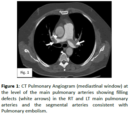
The images of the CT pulmonary angiogram showed multiple filling defects in the RT and LT main Pulmonary arteries, their segmental and sub segmental arteries which was consistent with pulmonary embolism (Figure 1).
Figure 1: CT Pulmonary Angiogram (mediastinal window) at the level of the main pulmonary arteries showing filling defects (white arrows) in the RT and LT main pulmonary arteries and the segmental arteries consistent with Pulmonary embolism.
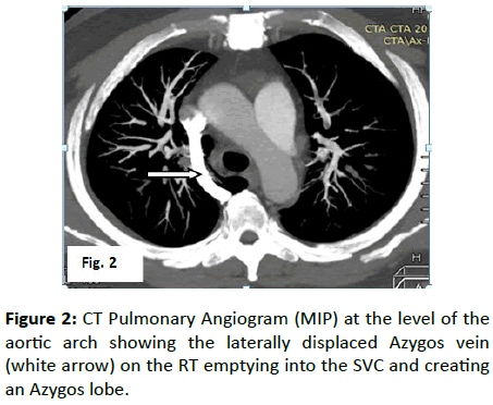
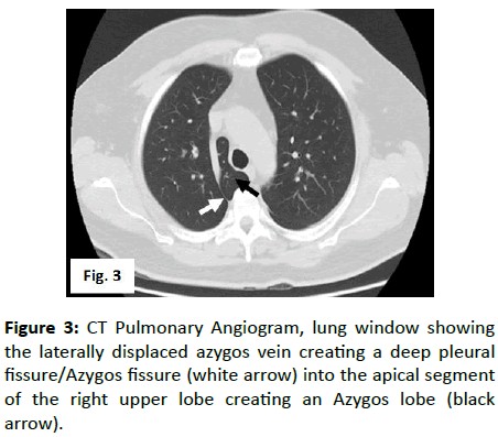
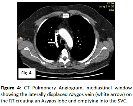
We also found incidentally a laterally displaced azygos vein creating a profound pleural crevice into the apical section of the correct upper projection creating an Azygos lobe and draining into the superior vena cava (SVC) (Figures 2-4). He was treated and later discharged by the neurosurgical unit and he is doing well.
Figure 2: CT Pulmonary Angiogram (MIP) at the level of the aortic arch showing the laterally displaced Azygos vein (white arrow) on the RT emptying into the SVC and creating an Azygos lobe.
Discussion
This case report describes a rare but significant variant anatomy of the right lung, found incidentally on a CT pulmonary angiogram in a 42-year old male presenting with pulmonary embolism. The worldwide incidence of Azygos lobe ranges between 0.2%-1.2% with approximately 0.4% seen on chest radiographs and 1% on anatomical dissections [5,6]. Azygos lobe is more common in males than females and there is a predilection for family inheritance [7,8].
Azygos lobe is in fact not a true accessory lobe as it is not an independent segment of the right lung [2,12]. It is rather a piece of the correct upper projection of the right lung as its arterial and bronchial supplies stem from the apical segment of the right upper lobe [6,13]. Though the left azygos lobe has been documented, it is extremely rare [14].
The azygos vein is ordinarily shaped by the association of the privilege subcostal vein and the correct rising lumbar vein at the degree of the L1/L2 vertebrae. It enters through the diaphragmatic aortic rest into the thoracic pit, rises along the anterolateral surface of the thoracic vertebrae, takes a bend at T4 and after that joins the predominant vena cava. The azygos vein curve shows up as a tear drop on x beam and is ordinarily situated at the caudal purpose of the privilege paratracheal stripe, at the privilege tracheobronchial edge. The azygos projection is framed when the privilege back cardinal vein, which is one of the forerunners of the azygos vein, infiltrates the correct lung pinnacle, instead of relocating over it. The cardinal vein conveys both pleural layers with it, bringing about ensnarement of a segment of the correct upper flap. The twofold creases of instinctive and parietal pleura structure a mesentery like structure, named the mesoazygos or azygos gap, containing the azygos vein curve in its lower generally portion [15].
Radiographicaly, the Azygos projection is normally very much observed on the chest radiograph, where it is restricted by the azygos crevice, a fine, raised (in respect to the mediastinum) line that crosses the zenith of the correct lung and on CT pneumonic Angiography demonstrates the profound infiltration of the lung behind the Superior Vena Cava (SVC) and the trachea. The azygos gap reaches out from the sidelong part of the vertebral body posteriorly, to the privilege brachiocephalic vein and SVC anteriorly. This comprises of two layers of parietal pleura and two layers of instinctive pleura. The azygos vein is viewed as a thicker structure following a similar way as the crevice and this was found in the pictures of our patient. The situation of the curve is higher than when it pursues an intramediastinal course. The azygos vein as a rule finishes in the SVC, yet sometimes in the privilege brachiocephalic vein. The azygos vein was seen finishing off with the SVC in our patient.
Two cases were accounted for where the phrenic nerve was seen coursing inside the azygos crevice. The azygos crevice or pleural folds helps in avoiding spread of the disease to the azygos projection from contiguous pieces of the lung. Nonetheless, numerous instances of unconstrained pneumothorax related with an azygous flap have been accounted for it. Intermittent hemoptysis, as a complexity of an azygos projection, has additionally been reported. An azygos vein aneurysm can likewise happen, which may present as a round or oval paratracheal shadow [15].
We could not obtain his Chest Radiograph and the scannogram obtained before the CT Pulmonary angiogram did not reveal much.
The first case of pulmonary azygos lobe reported in Ghana was encountered during a thoracotomy for modified Blalock- Tausig shunt in a 3-year old boy [16].
A detailed understanding or knowledge of this anatomical variant is crucial as azygos lobe might increment the danger of neurovascular wounds with resultant excessive blood loss or phrenic nerve injury and other complications during thoracoscopic procedures for sympathectomy or thoracic surgery and radiologists also need to know this anatomic variant in order not to mistaken it for a pulmonary pathology [17,18].
Conclusion
Radiologists should be aware of this rare anatomic variant, look for it and note it in their reports as knowledge of this variety is significant so as to counteract misdiagnosis and additionally pointless intercessions. In addition knowledge of the various lesions that might occur in the azygos lobe will cause early detection. Thoracic surgeons and Respiratory care physicians should also be aware of this anatomic variant and the clinical implications of azygos lobe as this can compromise the success of thoracic procedures such as endoscopic thoracic sympathectomy or lobectomy.
Conflicts of Interest
None
References
- Felson B (1989) The azygos lobe: its variation in health and disease. Seminars in Roentgenol 24: 56–66.
- Salve VT, Atram J S, Mhaske Y V (2015) Azygos lobe presenting as right para-tracheal shadow. Lung India 32: 85–86.
- Gill AJ, Cavanagh, SP, Gough, MJ (2004) The azygos lobe: An anatomical variant encountered during thoracoscopic sympathectomy. Eur J Vasc Endovasc Surg, 28: 223-224.
- Mata J, Caceres J, Alegret X, Coscojuela P, De Marcos J, et al. (1991) Imaging of the azygos lobe: Normal anatomy and variations. AJR Am J Roentgenol 156: 931-937.
- Arakawa T, Terashima T, Miki A (2008) A human case of an azygos lobe: Determining an anatomical basis for its therapeutic postural drainage. Clin Anat, 21: 524-530.
- Gourahari P, Satyajeet S, Siladitya M, Yera D, Suman KJ, et al. (2017) Azygos lobe-a rare anatomical variant. J Clin Diagn Res 11: 243-248.
- Shinichi Fukuhara, Marissa Montgomery, Angelo Reyes (2013) Robot-assisted azygos lobectomy for adenocarcinoma arising in an azygos lobe. Interact Cardiovasc Thorac Surg 16: 715–717.
- Fisher MS (1985) Re: Adam's lobe. Radiology 154: 547-547.
- Postmus PE, Kerstjens J M, Breed A, Jagt EVD (1986) A Family with Lobus Venae Azygos. Chest, 90: 298-299.
- Darlong LM, Ram D, Sharma A, Sharma AK, Iqbal SA, et al. (2017) The Azygous Lobe of the Lung. In: The Case of Lung Cancer. Indian J Surg Oncol, 8: 195-197.
- Kotov G, Dimitrova I N, Iliev A (2018) A Rare Case of an Azygos Lobe in the Right Lung of a 40-year-old Male. 10: 2780.
- Chabot-Naud A, Rakovich G, Chagnon K, Ouellette D, Beauchamp G, et al. (2011) A curious lobe. Can Respir J 18: 79-80.
- Wall J, Stawicki SP (2017) The azygos lobe. Int J Acad Med 3: 189-90.
- Lesser MB (1964) Left Azygos Lobe: Report of a case. Chest 46: 95-96.
- Jamal A Amos L, Kevin B Martin, Joel P (2018) Azygos lobe: A rare cause of right paratracheal opacity. Respir Med Case Rep 23: 136–137.
- Edwin F, Gyan B, Tettey M, Kotei D, Frimpong-Boateng K, et al. (2009) A pulmonary azygos lobe encountered during thoracotomy for modified Blalock-Taussig shunt.
- Delalieux S, Hendriks J, Valcke Y, Somville J, Lauwers P, et al. (2006) Superior sulcus tumor arising in an azygos lobe. Lung Cancer 54: 255-257.
- Ndiaye A, Ndiaye NB, Ndiaye A, Diop M, Ndoye JM, et al. (2012) The azygos lobe: an unusual anatomical observation with pathological and surgical implications. Anat Sci Int 87: 174-178.
Open Access Journals
- Aquaculture & Veterinary Science
- Chemistry & Chemical Sciences
- Clinical Sciences
- Engineering
- General Science
- Genetics & Molecular Biology
- Health Care & Nursing
- Immunology & Microbiology
- Materials Science
- Mathematics & Physics
- Medical Sciences
- Neurology & Psychiatry
- Oncology & Cancer Science
- Pharmaceutical Sciences


