Pelvis Arteriovenous Malformation in Males: A Case Study
Mostafa Shendy1* and Mohammed Dallash2
1 Department of Urology, Leighton Hospital, Middlewich Rd, Crewe CW1 4QJ, UK
2 Department of Urology, Wexham Hospital, Slough, UK
- *Corresponding Author:
- Mostafa Shendy
Department of Urology,
Leighton Hospital,
Middlewich Rd,
Crewe CW1 4QJ,
UK;
E-mail: Mostafa.shendy@nhs.net
Received Date: October 29, 2021; Accepted Date: November 12, 2021; Published Date: November 19, 2021
Citation: Shendy M and Dallash M (2021) Pelvis Arteriovenous Malformation in Males: A Case Study. J Nephrol Urol Vol.5 No.4:18.
Abstract
Pelvis Arteriovenous malformation is considered relatively rare and usually discovered accidentally as the patient comes complaining of some urinary symptoms (usually dysuria). I am reporting a case report of 60 years old male who was referred for visible hematuria less than 2 weeks referral pathway also complained of urinary tract infection like symptoms (dysuria).
Keywords
Pelvis arteriovenous malformation; Prostate abnormalities; Prostate cancer; Magnetic Resonance Imaging (MRI)
Introduction
A Midstream Specimen of Urine (MSU) was negative for infection, and during The digital rectal examination right lobe of the prostate felt hard, so the patient was investigated with Magnetic Resonance Imaging (MRI) prostate which gave the possibility of arteriovenous malformation, computerized tomography and us pelvis with doppler gave the diagnosis of thrombosed venous malformation around the prostate and pushing the bladder to the right side with no arterial involvement, urology team and the vascular team was involved and the decision was to treat prostatitis associated with it and repeat the scan after 6 months to confirm resolve prostatitis and completely thrombosed the venous malformation [1,2].
Case Study
A 60 years old male was referred via hematuria pathway due to visible hematuria, patient complained of Urinary Traction Infection (UTI) like symptoms. However no growth on MSU, physical examination was normal, Digital rectal examination showed 40-gram prostate and was hard on the right lobe, PSA 0.81 ug/l, flow rate showed Q max: 19.4 ml/s, Post voiding residual only 24 ml.
Medical history: Recent infection with Noro virus, inflammatory bowel disease, non-smoker, non-alcoholic.
Laboratory investigation
Showed serum creatinine 66 umol/L, EGFR>90, PSA 0.81 Ug/l HB: 122 WBCs: 4.4 Platelets: 272
Imaging investigation
Flexible cystoscopy: No abnormality was seen
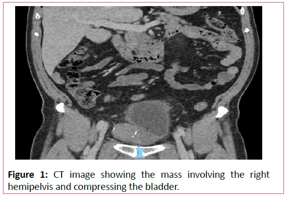
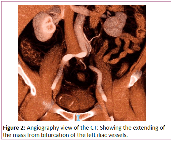
Clinical Teaching Unit (CTU) showed non enhancing mass lesion with eggshell calcification arising in the right hemipelvis extending from the bifurcation of the common iliac vessels anterior inferior involving the right side of the prostate and urinary bladder with a degree of compression of the right distal ureter but no obstruction (Figures 1 and 2) [3].
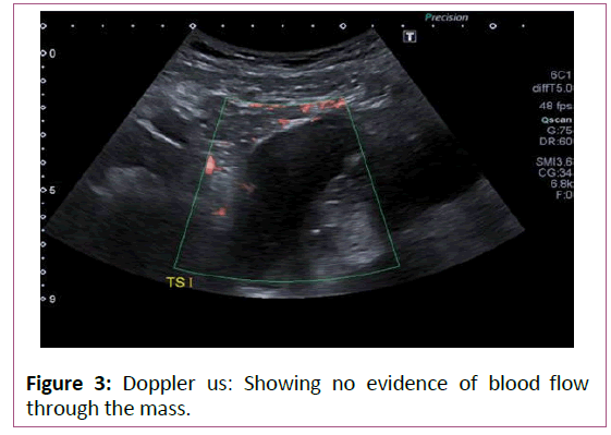
Transabdominal US pelvis: Well-defined serpiginous echo poor structure anteriorly within the right pelvis displacing the bladder to the left, the contents of this appear fluid in nature, and there is no evidence of any colour doppler flow (Figure 3) [4].
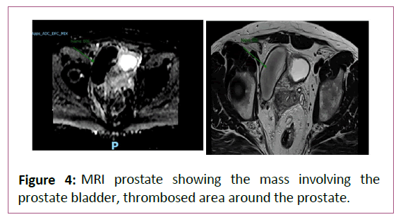
MRI prostate: Large right-sided serpiginous pelvic lesion wraps around, and distort the bladder neck, with evidence of the unusual appearance of large thrombosing right pelvic arterialvenous malformation, the structure appears to encase branches of the internal iliac artery but no connection between the artery and the structure, but some continuity between the structure and internal iliac vein which suggests low flow or thrombosed venous malformation (Figure 4)[5].
Diagnosis
The images were discussed in the X-ray meeting and the transvaginal ultrasound (US) doppler pelvis showed no blood flow with calcified edges which fits with long-standing thrombosed vascular malformation which most likely secondary to prostatitis needed to be treated, also recommended repeat of MRI after 6 months, refer to a vascular surgeon for an opinion [3,4].
The patient was referred for vascular surgery and he was discussed in The vascular Multidisciplinary Team (MDT) and after reviewing the imaging they came to conclusions that this is vascular malformation which is mainly venous with no arterial connection and it is better to be left without intervention unless a prostate biopsy is needed and in this case, he will need embolism the venous malformation before any prostate intervention [1,6].
Management
The patient was treated with triple therapy (co-trimoxazole, alpha-blocker, and ibuprofen).
Follow up with another MRI prostate after 6 months.
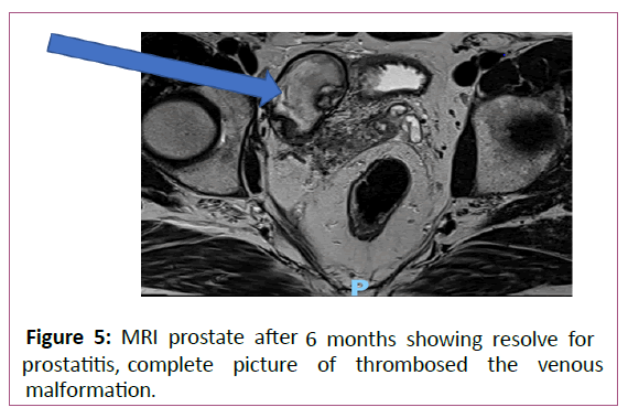
MRI prostate after six months showed
The prostate has reduced a little in size now measuring 42 cc and features of prostatitis seen previously have resolved and been Replaced by some low volume fibrosis [6]. The right seminal vesicle changes have matured; the large serpiginous presumed right pelvic Arteriovenous Malformations (AVM) has reduced a little, in size and the features of thrombosis have now matured and are complete.
It remains unclear whether the original prostatic inflammatory changes were reactive or the underlying cause for the thrombosis (Figure 5) [7].
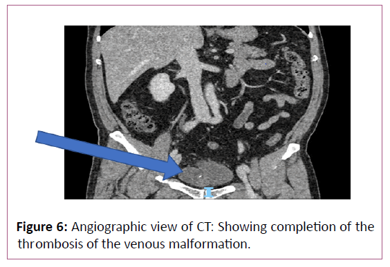
Computerized tomography abdomen pelvis with contrast showed
Serpiginous structure is once again seen on the right side of the pelvis, with no change in size. This demonstrates peripheral wall calcification with some peripheral wall enhancement particularly superiorly. The structure contains fluid with no enhancement [2,3]. Superiorly the structure appears to encase branches of the internal iliac artery but there is no definite connection between the artery and the structure. There does appear to be some continuity between the structures and is the internal iliac vein (Figure 6) [4-7].
Discussion
Pelvic AVM in males is a rare condition that could present with simple lower urinary tract symptoms, sometimes could be connected to the main artery like internal iliac artery needs embolization and could venous malformation which could be managed conservatively and regular follow-up [3-5].
The condition should be discussed in the MDT meeting before considering any intervention. The patient who comes with lower urinary symptoms should be evaluated in the best proper way, full medical history should be taken, full physical examination should be done including digital rectal examination, if any prostate suspicion area felt, patient, should have MRI prostate if it isn't contraindicated even if PSA is normal [7]. Visible hematuria should be taken seriously with full imaging of the upper urinary tract (US or CT urogram) and lower urinary tract imaging (flexible cystoscopy).
Conclusion
Prostate biopsy should be considered in the highly suspected prostate lesion But also shouldn't be rushed till it recommended by MRI, In case of prostatitis triple therapy (AB, alpha-blocker, anti-inflammatory medication) should be considered also treatment can take a long time for that repeat the imaging should be done at least after 6 months.
References
- Ledson MJ, Wahbi Z, Harris P (1999) A large pelvic arteriovenous malformation in an adult patient with cystic fibrosis. Postgrad Med J 75(884): 353-354.
- Zabicki B, Holstad MJV, Limphaibool N, Juszkat R (2019) Endovascular therapy of arteriovenous malformation in a male patient with severe post-coital pelvic pain. Pol J Radiol 27(1): e258-e261.
- Bekci T, Yucel S, Turgut E, Soylu AI (2015) Giant congenital pelvic AVM causing cardiac failure, fiplegia, and neurogenic bladder. Pol J Radiol 13(80): 388-390.
- Vos LD, Bom EP, Vroegindeweij D, Tielbeek AV (1995) Congenital pelvic arteriovenous malformation: A rare cause of sciatica. Clin Neurol Neurosurg 97(3): 229-232.
- Murakami K, Yamada T, Kumano R, Nakajima Y (2014) Pelvic arteriovenous malformation treated by transarterial glue embolisation combining proximal balloon occlusion and devascularisation of multiple feeding arteries. BMJ Case Rep 6(1): bcF013203492.
- Mallios A, Laurian C, Houbballah R, Gigou F, Marteau V, et al. (2011) Curative treatment of pelvic arteriovenous malformation–an alternative strategy: Transvenous intra-operative embolisation. Eur J Vasc Endovasc Surg 41(4): 548-553.
- Koganemaru M, Abe T, Iwamoto R, Suenaga M, Matsuoka K, et al. (2012) Pelvic arteriovenous malformation treated by superselective transcatheter venous and arterial embolization. Jpn J Radiol 30(6): 526-529.
Open Access Journals
- Aquaculture & Veterinary Science
- Chemistry & Chemical Sciences
- Clinical Sciences
- Engineering
- General Science
- Genetics & Molecular Biology
- Health Care & Nursing
- Immunology & Microbiology
- Materials Science
- Mathematics & Physics
- Medical Sciences
- Neurology & Psychiatry
- Oncology & Cancer Science
- Pharmaceutical Sciences
