ISSN : 2576-3938
Journal of Emergency and Internal Medicine
Initial Presentation of Pneumonia Mimics Hampton's Hump
Chung-Jui Fu* and Kai-Huang Lin
Department of Genetics, Michigan Technological University, Changhua, Taiwan.
- *Corresponding Author:
- Chung-Jui Fu, Department of Genetics, Michigan Technological University, Changhua, Taiwan, Tel: 0932345845; E-mail: kaihuang@cch.org.tw
Received date: February 07, 2022, Manuscript No. IPJEIM-22-12606; Editor assigned date: February 10, 2022, PreQC No. IPJEIM-22-12606 (PQ); Reviewed date: February 25, 2022, QC No. IPJEIM-22-12606; Revised date: April 11, 2022, Manuscript No. IPJEIM-22-12606 (R); Published date: April 19, 2 022, DOI: 10.36648/2576-3938.6.4.001
Citation: Fu CJ, Lin KH (2022) Initial Presentation of Pneumonia Mimics Hampton’s Hump. J Emerg Intern Med Vol:6 No:4
Case Description
A 75-year-old man, who was admitted for hepatic arterial infusion chemotherapy, had previously undergone a total right lobectomy and cholecystectomy for a hepatocellular carcinoma. He completed four cycles of trans-arterial chemo-embolization and was started on targeted therapy with lenvatinib 4 months ago. He presented with chest pain after a 6-day-course of intra-arterial chemotherapy with cisplatin, mitomycin, and fluorouracil. His heart rate was 100 bpm and his oxygen saturation level was 100% on room air. On physical examination, he did not display signs of fever, productive cough, palpitation, diaphoresis, or dyspnea.
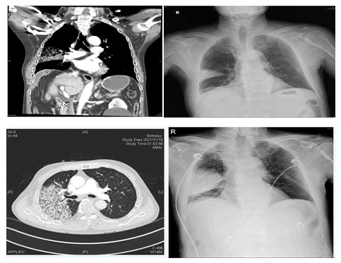
His condition rapidly deteriorated, and he developed hemoptysis and intolerable pleuritic pain 3 hours later. His oxygen saturation level dropped to 88% on room air, and oxygen therapy with Venturi mask (15 L/min) was initiated. Follow-up blood gas analysis showed hypoxia and elevated D-dimer levels (>10000 mg/mL) with unremarkable white blood cell count, troponin levels, and renal function. Chest radiography performed 4 hours later revealed rapid progression of the dome-shaped opacity with emphysematous change and a bulging fissure sign Computed tomographic pulmonary angiography revealed the presence of large areas of airspace consolidations and ground-glass opacities in the right lower lobe and right upper lobe. No filling defects in pulmonary arteries were noted.
A diagnosis of pneumonia by Klebsiella pneumoniae was confirmed via sputum culture analysis. The patient was started on antibiotic therapy according to antibiotic sensitivity report with a plan of pulmonary rehabilitation therapy. He was successfully extubated on the 34th day of admission (Figure 1).
References
- Nugent K, Moll J (2014) The Hampton hump in acute pulmonary embolism. J Emerg Med. 46:828-829.
[Crossref] [Google Scholar] [Indexed]
- Kawasaki MC, Mizumoto J (2022) Hampton's hump: hypoxia with lung consolidation mimicking pneumonia. JMA J. 5:135-136.
[Crossref] [Google Scholar] [Indexed]
Open Access Journals
- Aquaculture & Veterinary Science
- Chemistry & Chemical Sciences
- Clinical Sciences
- Engineering
- General Science
- Genetics & Molecular Biology
- Health Care & Nursing
- Immunology & Microbiology
- Materials Science
- Mathematics & Physics
- Medical Sciences
- Neurology & Psychiatry
- Oncology & Cancer Science
- Pharmaceutical Sciences

