Histomorphometric Effects of Crude Methanolic Extract of Garcinia kola Seeds on the Hippocampus of Adult Wistar Rats
Akinola BK*, Olawuyi TS and Sanusi T Nabeelat
Department of Human Anatomy, Federal University of Technology Akure, Akure, Ondo State, Nigeria
- *Corresponding Author:
- Akinola BK
Department of Human Anatomy,
Federal University of Technology Akure,
Akure, Ondo State,
Nigeria
Tel: 08032073693;
E-mail: bkakinola@futa.edu.ng
Received Date: October 29, 2021; Accepted Date: November 12, 2021; Published Date: November 19, 2021
Citation: Akinola BK, Olawuyi TS, Nabeelat ST (2021) Histomorphometric Effects of Crude Methanolic Extract of garcinia kola Seeds on the Hippocampus of Adult Wistar Rats. J Anat Sci Res Vol.4 No.5:5.
Abstract
This study was designed to investigate the histomorphometric effect of garcinia kola on nickel chloride induced neurotoxicity in adult Wister rats. Thirty (30) rats were used for this study. The rats were divided into five (5) groups of six (6) rats each. Group A received 1 ml/kg body weight distilled water, Group B received 20 mg/kg body weight of nickel chloride, Group C received 20 mg/kg body weight of nickel chloride and 100 mg/kg of crude extract of garcinia kola seeds, Group D received 20 mg/kg body weight of nickel chloride and 200 mg/kg of crude extract of garcinia kola seeds and Group E received 20 mg/kg of crude extract of garcinia kola seeds concomitantly for 21 days. At the end of the experimental period, brain tissues were harvested and taken for histopathological examination. Results obtained showed that the exposure of rats to nickel chloride lead to the degeneration of neuronal cells and significant loss of body weight. However, the administration of the crude extract of garcinia kola with nickel chloride limited the damaging effect of nickel chloride. The present findings showed the ameloriative potential of garcinia kola on nickel chloride induced neurotoxicity in adult wistar rat.
Keywords
garcinia kola; Hippocampus; Neurotoxicity; Nickel chloride; Jaundice
Introduction
garcinia kola among a few of the many is a medicinal plant found in Africa. It is an herb grown in Nigeria and usually has a bitter and resinous taste. It is called Bitter Kola in Nigeria. Other local Nigerian names include; Orogbo in Yoruba, Ugolu in Ibo and Akan in Urhobo [1]. garcinia kola has been referred to as a “wonder plant” because every part of it has been found to be of medicinal importance [2]. Studies have reported the use of this plant for the treatment of jaundice, high fever, and purgative and chewing stick. The plant also found usefulness in the treatment of stomach ache and gastritis [3]. The growing trees and the roots provide chew-sticks. The seeds of G. kola are edible and are traditionally served to visitors as an adjunct to the real colas (Cola nitida and Cola acuminata) [4].
Nickel and its compounds are known for their carcinogenic and toxic activities in human beings and experimental animals [5]. It is used as a trace metal for many animal species, microorganisms and plants, and therefore deficiency or toxicity symptoms may occur when little or much nickel is taken up [6]. Nickel chloride (Nickel (II) chloride), NiCl2, which is a compound of nickel is a crystalline inorganic compound that generate toxic gases upon heating. Overexposure to this compound can result to inflammation of the skin, damage of the lungs, kidneys, gastrointestinal tract and neurological system. The hippocampus has been a source of fascination from the very origins of neuroscience as a modern discipline. The complexity of this phylogenetically ancient structure, at both the gross and cellular level has encouraged study by generations of anatomists [7]. The hippocampus is a major component of the brain of humans and other mammals. It belongs to the limbic system and plays important role in long-term memory and spatial navigation [1]. Hence, this research study the effects of methanolic crude extract of garcinia kola seeds on nickel chloride induced hippocampal toxicity.
Materials and Methods
Plant material and authentication
The seeds of garcinia kola were purchased from Akure Market in Ondo State, Nigeria. Authentication of the seeds was carried out in the department of Crop Science and Production, Federal University of Technology Akure, with the voucher specimen number (0309).
Preparation of garcinia kola extract
The seeds were peeled and air dried for a period of five days. Then, the seeds were grinded into a powdered form and were also allowed to dry. 500 g of the powdered garcinia kola seeds was measured and soaked in 2250 ml of methanol for 72 hours with mixing at intervals. Afterwards, it was then filtered using a What man No 1 filter paper. The filtrate was allowed to evaporate to give the required brownishblack residue. The brownish-black residue was kept at 4oC for use.
Experimental animals
For the purpose of this study, 30 adult female and male Wister rats with weight ranging from 90 g-160 g was purchased. The animals would be kept in clean cages under standard temperature conditions (natural day/night cycle, ambient temperature) and allowed access to food and water adlibitum. All animals will be handled according to the National Institute of Health guidelines for the care and use of laboratory animals. The animals were acclimatized for 4 weeks prior to experiment.
Experimental design
The rats were randomly assigned into five (5) groups (A, B, C, D and E) with six animals per group to ensure even distribution of mean body weight across groups. The body weight and the average weight of each group were taken and recorded every two days.
The animals were treated as follows;
Group A received 1 ml/kg body weight (bwt) of distilled water for 4 weeks.
Group B received 20 mg/kg (bwt) of nickel chloride once daily for 4 weeks.
Group C received 20 mg/kg (bwt) of nickel chloride and 100 mg/kg of crude methanolic extract of garcinia kola seeds once daily for 4 weeks.
Group D received 20 mg/kg (bwt) of nickel chloride and 200 mg/kg of crude methanolic extract of garcinia kola seeds once daily for 4 weeks.
Group E received 20 mg/kg (bwt) of methanolic extract once daily for 4 weeks.
Animal sacrifice and sample collection
The rats were weighed and sacrificed 24 hours after the last dose treatment by cervical dislocation. The brain was harvested and weighed. After physical examination, the brains obtained were fixed in a labeled bottle with neutral buffered formalin.
Histological analysis
The brains were fixed immediately in 10% formalin for 48 hours. It was processed in paraffin sections. Blocks embedded in paraffin were cut into different sections (5 mm thickness) with a microtome. Sections were stained with hematoxylin and eosin (H and E). The stained sections were mounted with DPX and were observed under a microscope. Their photomicrograph was taken at varying magnifications with a Digital Camera (Image J). Histological evaluation was presented with tissue micrograph H and E x40, x100 and x400.
Results
Statistical analysis
All data obtained were subjected to statistical analysis. Values were expressed as Mean ± Standard Error of Mean (SEM) while one way ANOVA was used to test for the differences between treatment groups using Graph pad Prism. The results were considered significant at p values less than 0.05 (p<0.05). These were then presented with suitable tables and chart (Figures 1-8).
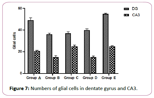
Figure 7: Numbers of glial cells in dentate gyrus and CA3.
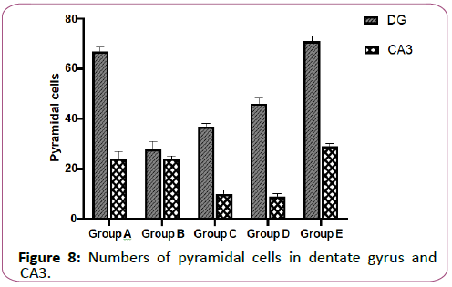
Figure 8: Numbers of pyramidal cells in dentate gyrus and CA3.
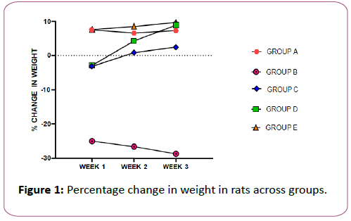
A line graph showing the change in body weight of rats across groups, Results are represented as Mean ± Standard Error of Mean (SEM), n=6 rats in each group. Value of p ˂ 0.05 was considered significant.
Effects of garcinia kola on the hippocampus across groups
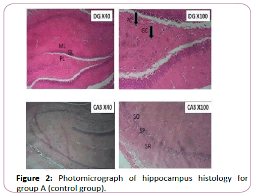
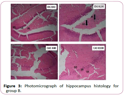
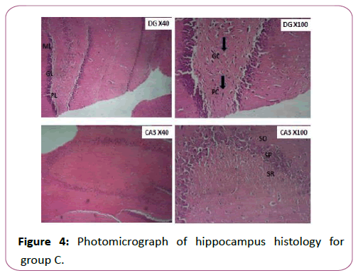
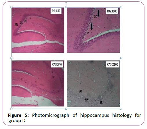
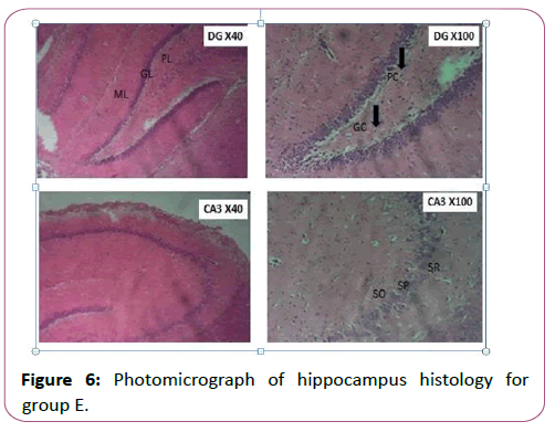
The results of the histology were shown in Figures 2-6 as mentioned below. The photomicrograph of the histological sections across groups revealed that the adminstration of garcinia kola extract ameliorates the histoachitecture of the hippocampus of group C, D and E when compared to group B. The observation of the control group A showed normal hippocampal morphology. This is in complete contrast to group B which showed structural disorganization typical of hippocampal damage.
Figure 2 showing normal histologic features of 2 areas of the hippocampus (DG and CA3). CA: Cornu Ammonis; DG: Dentate Gyrus; Hi-Hilus of DG; SO: Stratum Oriens; SP: Stratum Pyramidale (Pyramidal layer); SR: Stratum Radiatum; PC: Pyramidal cell; GC: Glial cell; ML: Molecular layer; PL: Polymorphiclayer and GL: Granular layer H and E (Magnification x 40 and x 100).
Figure 3 showing distorted pryramidal cells and glial cells which indicate cell loss ( Neurodegeneration) in 2 areas of the hippocampus (DG and CA3). CA: Cornu Ammonis; DG: Dentate Gyrus; Hi: Hilus of DG; SO: Stratum Oriens; SP: Stratum Pyramidale (Pyramidal layer); SR: Stratum Radiatum; PC: Pyramidal cell; GC: Glial cell; ML: Molecular layer; PL: Polymorphiclayer and GL: Granular layer. H and E; (Magnification x40 and x100).
Figure 4 showing signs of recovery in the neurons in 2 areas of the hippocampus (DG and CA3).CA: Cornu Ammonis; DG: Dentate Gyrus; Hi: Hilus of DG; SO: Stratum Oriens; SP: Stratum Pyramidale (Pyramidal layer); SR: Stratum Radiatum; PC: Pyramidal cell; GC: Glial cell; ML: Molecular layer; PL: Polymorphiclayer and GL: Granular layer H and E; (Magnification x40 and x100).
Figure 5 showing more improved signs of recovery in the neurons in 2 areas of the hippocampus (DG and CA3) when compared to group C. CA: Cornu Ammonis; DG: Dentate Gyrus; Hi: Hilus of DG; SO: Stratum Oriens; SP: Stratum Pyramidale (Pyramidal layer); SR: Stratum Radiatum; PC: Pyramidal cell; GC: Glial cell; ML: Molecular layer; PL: Polymorphic layer and GL: Granular layer H and E; (Magnification x40 and x100).
Figure 6 showing normal histologic features in 2 areas of the hippocampus (DG and CA3) like that of group A. CA: Cornu Ammonis; DG: Dentate Gyrus; Hi: Hilus of DG; SO: Stratum Oriens; SP: Stratum Pyramidale (Pyramidal layer); SR: Stratum Radiatum; PC: Pyramidal cell; GC: Glial cell; ML: Molecular layer; PL: Polymorphiclayer and GL-Granular layer H and E (Magnification x40 and x100).
Effects of garcinia kola on neuronal cell count of the hippocampus of an adult wistar rats
Here Figures 7 and 8 showing the effect of garcinia kola on neuronal cell count of the hippocampus of an adult wistar rats.
Each value represented Mean ± SEM, n=6 readings. Value of p˂0.05 was considered significant.
Each value represented Mean ± SEM, n=6 readings. Value of p˂0.05 was considered significant.
Discussion
Heavy metal pollution is a global problem due to its persistent accumulation in the environment. Chronic exposure to heavy metals could affect mental health. Among these metals is nickel (Ni), a toxic transition metal used in industry and found in air, water, and food [8]. Human overexposure to Ni and the increasing application of Ni compounds in modern technologies is a major public health issue. It has been reported that Ni exposure causes harmful effects in living organisms, such as genotoxicity, teratogenicity, immunotoxicity, and carcinogenicity [9].
This present study showed that administration of nickel chloride resulted in percentage loss of body weight in group B when compared with group A. Samir and Vine (2013) reported that animals treated with nickel chloride showed obvious significant decrease (p <0.001) in body weight gain as compared to control group. Gathwan also reported significant decrease in body weight of mice following nickel chloride administration at moderate and high doses [10]. The effect of nickel chloride has been recognized as strong biological poison (bio reductant) because of its destructive nature and tendency to accumulate in organisms and to undergo amplification. garcinia kola extract enhanced increase in percentage weight gain of rats in group C and D when juxtaposed with group A. In group E, significant body weight gain was observed. The observation is in accord with Adaramoye who reported that garcinia kola significantly increased the body weight of rat [11]. Adesuyi investigated the nutritional and phytochemical component of garcinia kola [12]. He reported that garcinia kola is a good source of proteins; tannin, oxalate etc. and carbohydrates. This may explain why animals treated with GK only had significant weight gain.
The administration of nickel chloride showed destructive properties. Unlike group A, C, D and E, group B showed higher destruction of cell in the Dentate Gyrus and CA3 which clearly shows that nickel chloride induces neurotoxicity in the brain. Ahmed affirmed the neurotoxic properties of nickel [13-15]. He observed that Ni induced demyelination in limited areas in varying degrees and necrotic changes in the brain. This is because heavy metals have been shown to induce neurotoxicity and subsequent behavioral effects by the generation of reactive oxygen species (ROS) from increased extracellular dopamine [13]. The hippocampal neurons of group C and D showed better histological and morphological integrity than group B. This finding was in agreement with that obtained by Ijomone [1]. He found that garcinia kola affords protection to hippocampal neurons of rats following the administration of methamephline. Omotoso studied the effects of garcinia kola on hippocampus of rats after the administration of cuprizone [14]. He affirmed the destructive properties of cuprizone as well as the protective effect of garcinia kola on rat. In his research, cellular clusters with pyknotic characteristics were observed in the CA3 region and poorly stained granule cells in the granular layer of the dentate gyrus of hippocampus of cuprizone treated mice. The garcinia kola then limited the damaging effects of cuprizone on the morphology and functions of hippocampus in mouse and also increased the cellular structure of hippocampus which was also observed in GROUP E. The protection afforded by Garicinia kola is probably due to its antioxidant ability. It is also probable that the level of protection afforded in this study could have been highly improved if garcinia kola was given for a longer duration and/or at higher dose than that used in this study.
Neuronal count denotes that the histology of GROUP A is well structured and intact. This is because the pyramidal and glial cells were much as expected. That of GROUP B reduced drastically due to the neurodegeneration caused by nickel chloride. Following the administration of garcinia kola at 100 and 200 mg/kg, the no of pyramidal and glial cells increased. This shows the protective ability of this wonderful herb. Apart from its ability to reduce the rate of neurodegeneration, garcinia kola administration also increased the no of cells in GROUP E when compared to the normal histology of GROUP A.
Conclusion
Based on the data of this study, it was observed that the crude methalonic extract of garcinia kola was very potent in preventing and protecting rat model from nickel chloride induced neurotoxicity. It also further established the assertion that nickel chloride decreases body weight of rats at higher doses at the same time causing hippocampal damage.
Recommendation
Water generally, contains Ni at concentrations ˂10 μg/L. Ni concentrations in highly contaminated fresh waters may reach as high as several hundred to 1000 μg/L. World health organization’s together with NAFDAC should focus on the contents of water produced in industries. Further investigation is necessary to shed light on the mechanism of neurotoxicity caused by nickel chloride and the protective effects on garcinia kola on the structures that makes up the limbic system following nickel chloride toxicity.
References
- Ijomone o M (2011) Protective effects of kolaviron, an extract of garcinia kola, on the hippocampal neurons of wistar rats exposed to methamphetamine.
- Adegboye MF, Akinpelu DA, Okoh A I (2008) The bioactive and phytochemical properties of garcinia kola (Heckel) seed extract on some pathogens. Afr. J. Biotechnol 7(21): 3934-3938.
- Iwu M M, Igboko O A, Okunji C O, Tempesta M S (1990) Antidiabetic and aldose reductase activities of biflavanones of garcinia kola. J Pharm Pharmacol 42(4): 290-292.
- Madubunyi I I (1995) Antimicrobial activities of the constituents of garcinia kola seeds. Int j pharmacogn 33(3): 232-237.
- Das K K, Das S N, Dhundasi S A (2008) Nickel, its adverse health effects and oxidative stress. Indian J Med Res 128(4): 412.
- Cempel M, Nikel G (2006) Nickel: a review of its sources and environmental toxicology. Pol J Environ Stud 15(3): 375–382.
- Thammaroj J, Santosh C, Bhattacharya J J (2005) The hippocampus: modern imaging of its anatomy and pathology. Pract. Neurol 5(3): 150-159.
- Bahadir Z, Ozdes D, Bulut V N, Duran C, Elvan H, et al. (2013) Cadmium and nickel determinations in some food and water samples by the combination of carrier element-free coprecipitation and flame atomic absorption spectrometry. Toxicol Environ Chem 95(5): 737-746.
- Cameron K S, Buchner V, Tchounwou P B (2011) Exploring the molecular mechanisms of nickel-induced genotoxicity and carcinogenicity: a literature review. Rev Environ Health 26(2):81-92.
- Gathwan K H, Al-Karkhi I H T, AL-Mulla E A J (2013) Hepatic toxicity of nickel chloride in mice. Res. Chem. Intermed 39(6): 2537-2542.
- Adaramoye OA, Nwaneri VO, Anyanwu KC, Farombi EO, Emerole, GO (2005) Possible anti‐atherogenic effect of kolaviron (a garcinia kola seed extract) in hypercholesterolaemic rats. Clin Exp Pharmacol Physiol 32(1‐2): 40-46.
- Adesuyi AO, Elumm IK, Adaramola FB, Nwokocha AGM (2012) Nutritional and phytochemical screening of garcinia kola. Adv. J. Sci. Technol 4(1): 9-14.
- Ahmed M K, Islam M S, Habibullah-Al-Mamun M,Hoque M F (2015) Preliminary assessment of heavy metal contamination in surface sediments from a river in Bangladesh. Environ Earth Sci 73(4): 1837-1848.
- Omotoso G O, Olajide O J, Gbadamosi I T, Adebayo J O, Enaibe B U, et al. (2019) Cuprizone toxicity and garcinia kola biflavonoid complex activity on hippocampal morphology and neurobehaviour. Heliyon 5(7): e02102.
- Eisler R (1998) Nickel hazards to fish, wildlife, and invertebrates: a synoptic review. US Department of the Interior, US Geological Survey, Patuxent Wildlife Research Center.
Open Access Journals
- Aquaculture & Veterinary Science
- Chemistry & Chemical Sciences
- Clinical Sciences
- Engineering
- General Science
- Genetics & Molecular Biology
- Health Care & Nursing
- Immunology & Microbiology
- Materials Science
- Mathematics & Physics
- Medical Sciences
- Neurology & Psychiatry
- Oncology & Cancer Science
- Pharmaceutical Sciences
