Femoral Length Reconstruction in Adults: An Osteometric and Radiographical Approach Using Regression Equations
Sunday Elijah1*, Akpan Ekanem2 and Aniekan Peter2
1Department of Human Anatomy, PAMO University of Medical Sciences, Port- Harcourt, Nigeria
2Department of Human Anatomy, University of Uyo, Nigeria
- *Corresponding Author:
- Sunday Elijah
Department of Human Anatomy, Faculty of Basic Medical Sciences, PAMO University of Medical Sciences, Port- Harcourt, Nigeria
Tel: +2348036221623
E-mail: elijahsunday85@gmail.com
Received Date: May 19, 2021; Accepted Date: June 02, 2021; Published Date: June 09, 2021
Citation: Elijah S, Ekanem A, Peter A (2021) Femoral Length Reconstruction In Adults: An Osteometric And Radiographical Approach Using Regression Equations. J Anat Sci Res Vol.4 No.3
Abstract
The increase in extremist mass killings and fatal automobile crash has greatly multiplied the rate at which forensic anatomists and anthropologists are faced with dismembered and mixed up body remains. This study was undertaken to reconstruct femoral length from its landmarks using radiological and anthropometric parameters. 600 bones and 600 radiographs were measured using an anthropometric board, an anthropometric tape and digital caliper and on the radiograph, a transparent ruler was used. The femoral maximum length; femoral proximal breadth; anterior posterior neck diameter; vertical head diameter; medial-lateral subtrochanteric diameter with gluteal tuberosity; subtrochanteric circumference with gluteal tuberosity; anterior-posterior subtrochanteric diameter with gluteal tuberosity; anterior-posterior diameter at mid-shaft; mediallateral mid-shaft diameter; femoral mid-shaft circumference; femoral epicondylar breadth and femoral bicondylar breadth were measured. No significant difference in the mean value was found between bones and radiographs although males showed higher mean length compared to females. Best predictors of length were femur proximal breath and vertical head diameter.
Keywords
Femur; Cartilage; Reconstruction; Regression; Anthropometric
Introduction
Where mass fatalities occur as a result of man-made or natural disasters, the estimation of stature is an important and reliable step in the process of identifying an individual during forensic investigation. Stature is said to vary within populations as a result of genetic combination, diet or social status [1]. Therefore the use of one monogram in another population may be a source of error in its estimate [2]. Stature has been successfully estimated from most long bones of the human body with relative ease and accuracy because it correlates with these bones [3,4].
The length of long bones has been estimated from anatomical landmarks on the bone using skeletal remains [5]. The length of lower limb bones plays important role in estimation of height of an individual hence most predictive formulas are based on the length of tibia and femur [6]. The present work aimed at ascertaining if landmarks on the x-ray radiographs of the femur correlated with the length of the bone, if these landmarks can be used to estimate the length of the bone, if the x-ray radiographs can be used in place of the bone or in combination with the bone to estimate the ante-mortem stature of an individual in a forensic investigation.
Materials and Methods
Six hundred femora pooled from Anatomical Museums and X-ray radiographs from hospitals within the Northeast, Northwest, North-central, Southeast, Southwest and South-south of Nigeria were utilized. All samples were assessed to eliminate bones with obvious pathological damages or inabilities to locate and identify landmarks. Radiographs used were the ones that showed the entire length of the bone with sharp image in the anteriorposterior view and with no case of trauma.
On bony samples, a digital vernier caliper calibrated to 0.1 mm was used for measuring dimension; an anthropometric board calibrated to 0.1 cm was used for taking full length measurement and an anthropometric tape calibrated to 0.1 cm was used for taking circumferential measurement; while on the x-ray radiographs, a transparent ruler calibrated to 0.1 cm was used for all measurements taken. Bones collected were identified and separated into right and left. Radiograph samples were separated as either belonging to male or female and then into rights and left. The landmarks used in the study were defined as follows [5- 8].
Femoral measurements
The rats were randomly divided into seven groups of ten animals each
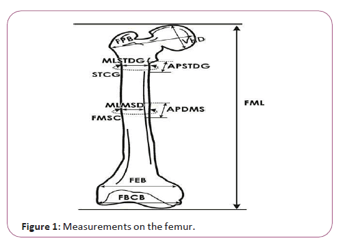
1. Measurement of the femoral maximum length (FML) was measured from the most superior point on the head of the femur to the most inferior point on the distal-medial condyle.
2. Femoral proximal breadth (FPB) was measured from the head of the femur to the greater trochanter.
3. Anterior posterior neck diameter (APND) was measured as the anterior-posterior diameter of the neck of femur.
4. Vertical head diameter (VHD) was measured as the horizontal diameter of the head of femur.
5. Medial-lateral subtrochanteric diameter with gluteal tuberosity (MLSTDG) was taken as the medial-lateral diameter measured at the point of greatest lateral expansion of the femur inferior to the lesser trochanter including the gluteal tuberosity.
6. Subtrochanteric circumference with gluteal tuberosity (STCG) was taken as the circumference measured on the shaft inferior to the lesser trochanter at the same level of the sagittal and transverse subtrochanteric diameters including the gluteal tuberosity.
7. Anterior-posterior subtrochanteric diameter with gluteal tuberosity (APSTDG) was taken as the anterior posterior diameter measured at the point of greatest lateral expansion of the femur below the lesser trochanter including the gluteal tuberosity.
8. Anterior-posterior diameter at mid-shaft (APDMS) was taken as the anterior-posterior diameter measured approximately at the midpoint of the diaphysis, at the highest elevation of the linea aspera. This measurement is perpendicular to the ventral surface.
9. Medial-lateral mid-shaft diameter (MLMSD) was measured at right angles to the anterior posterior diameter of the mid-shaft. The linea aspera should be midway between the two arms of the caliper.
10. Femoral mid-shaft circumference (FMSC) was measured at the mid-shaft at the same level of the sagittal and transverse diameters.
11. Femoral epicondylar breadth (FEB) was measured as the maximum distance from the most lateral point on the lateral epicondyle to the most medial point on the medial epicondyle.
12. Anterior-posterior diameter of the medial condyle (APDMC) was measured as the distance between the most posterior point on the medial condyle and lip of the patellar surface perpendicular to the axis of the shaft.
13. Measurement of Femoral bicondylar breadth (FBCB) was measured from the most lateral and posterior projection of the lateral condyle, to the most medial and posterior projection of the medial condyle (Figure 1).
Measurements on the radiograph of femur
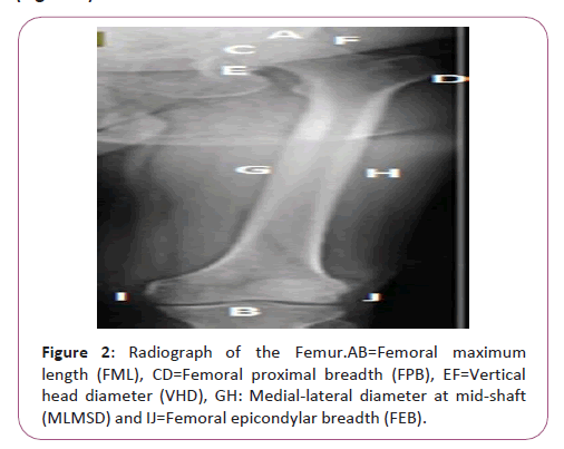
1. Femoral maximum length (FML) was measured as the distance from the most superior point on the head of the femur to the most inferior point on the distal-medial condyle.
2. Femoral proximal breadth (FPB) was measured as the width from the head of the femur to the greater trochanter.
3. Vertical head diameter (VHD) was measured as the diameter of the head of femur taken in the horizontal plane.
4. Medial-lateral diameter at mid-shaft (MLMSD) was measured as the distance from the medial to the lateral aspect of the midshaft.
5. Femoral epicondylar breadth (FEB) was measured as the maximum distance from the most lateral point on the lateral epicondyle to the most medial point on the medial epicondyle (Figure 2).
Statistical analysis
To eliminate bias, the same measurements were verified from 30 randomly selected samples by two evaluators, the examiner and the recorder using the same unit and instrument and technical error of measurements were calculated. The intraand inter- observer technical error of measurement (TEM) was calculated using [TEM={√ΣD2/2N}, where D=difference between the measurements, N=number of samples measured] and the coefficient of reliability was also calculated using [R={1- (TEM)2/SD2} where SD=standard deviation of all measurements] [9,10]. The mean, standard deviation, minimum, maximum and standard error were determined. Comparisons between the right and left variables were performed using student’s t-test. Pearson’s correlation coefficient was carried out to assess the relationship between the variables (independent variable, x) and length (ML – dependent variable, y). Regression analysis was undertaken to find the variables that related to length and for estimating length using equations. Regression equations were derived to construct the length of each bone from the significant variables. Simple regression models at y=mx +c were derived, where ‘c’ is a constant, ‘m’ is the regression coefficient and the asterisk “*” denotes significant values at p<0.05. After excluding highly correlated variables using a stepwise method, multivariate regression equations were derived and the most suitable parameter for predicting length was determined using the highly correlated variables. Analysis was done using SPSS (version 21) statistical package.
Ethical clearance
Compliance with institutional rules with respect to human experimental research and ethics was strictly adhered to in the course of this study. Written approval was obtained from the Human Research Ethics committee with reference number FCT/ UATH/HREC/1085.
Results
The technical error of measurement (TEM) for the femur and its radiographs showed coefficient of reliability, R>0.95 in all cases, therefore the measurements were regarded as reliable (Table 1).
| Intra-observer error | Inter-observer error | ||||||||
|---|---|---|---|---|---|---|---|---|---|
| S/N | Variable | TEM (b) | (r) | R (b) | (r) | TEM (b) | (r) | R (b) | R (r) |
| 1 | FML | 0.693 | 0.699 | 0.98 | 0.98 | 0.694 | 0.699 | 0.98 | 0.98 |
| 2 | FPB | 0.17 | 0.179 | 0.98 | 0.98 | 0.439 | 0.179 | 0.98 | 0.98 |
| 3 | APND | 0.327 | - | 0.98 | - | 0.063 | - | 0.98 | - |
| 4 | VHD | 0.071 | 0.063 | 0.98 | 0.98 | 0.063 | 0.063 | 0.99 | 0.98 |
| 5 | MLSTDG | 0.071 | - | 0.98 | - | 0.063 | - | 0.98 | - |
| 6 | STCG | 0.063 | - | 0.98 | - | 0.134 | - | 0.98 | - |
| 7 | APSTDG | 0.13 | - | 0.98 | - | 0.045 | - | 0.98 | - |
| 8 | APDMS | 0.055 | - | 0.98 | - | 0.077 | - | 0.98 | - |
| 9 | MLDMS | 0.071 | 0.071 | 0.98 | 0.98 | 0.063 | 0.071 | 0.98 | 0.98 |
| 10 | FMSC | 0.195 | - | 0.98 | - | 0.2 | - | 0.98 | - |
| 11 | FEB | 0.141 | 0.176 | 0.98 | 0.98 | 0.164 | 0.176 | 0.98 | 0.98 |
| 12 | APDMC | 0.126 | - | 0.98 | - | 0.126 | - | 0.98 | - |
| 13 | FBCB | 0.148 | - | 0.98 | - | 0.161 | - | 0.98 | - |
TEM=Technical error of measurement; R=coefficient of reliability; (b)=bones; (r)=radiographs; Unit=cm; Number of samples=30
Table 1: Technical error for the measured parameters of femur using bones and radiographs parameters.
The mean length of the femur using bones: The mean length of the femur was 46.56 ± 3.01 cm and 46.76 ± 3.23 cm for the right and left respectively. When the right and left femora were combined, the mean length was 46.66 ± 3.12 cm. No statistical significant difference in the mean length between the right, left and the combined femora was observed and all the variables correlated significantly with the length of femur (Table 2).
|
Right N=300 |
Left N=300 |
Combined N=600 |
||||||||||||||
|---|---|---|---|---|---|---|---|---|---|---|---|---|---|---|---|---|
| S/N | Variable | C | SE | Mean ± SD | M | P value | C | SE | Mean ± SD | M | P value | C | SE | Mean ± SD | M | P-value |
| 1 | FPB | 33.72 | 0.05 | 8.93 ± 0.86 | 1.44 | 0.000* | 19.74 | 0.04 | 9.04 ± 0.71 | 2.99 | 0.000* | 28.1 | 0.03 | 8.98 ± 0.79 | 2.07 | 0.000* |
| 2 | APND | 39.43 | 0.02 | 2.64 ± 0.28 | 2.7 | 0.000* | 36.18 | 0.02 | 2.63 ± 0.31 | 4.02 | 0.000* | 37.63 | 0.01 | 2.64 ± 0.29 | 3.43 | 0.000* |
| 3 | VHD | 25.47 | 0.02 | 4.44 ± 0.34 | 4.75 | 0.000* | 22.71 | 0.02 | 4.47 ± 0.37 | 5.39 | 0.000* | 23.98 | 0.02 | 4.45 ± 0.35 | 5.09 | 0.000* |
| 4 | MLSTDG | 36.81 | 0.02 | 3.00 ± 0.31 | 3.24 | 0.000* | 33.89 | 0.02 | 3.11 ± 0.29 | 4.15 | 0.000* | 35.58 | 0.01 | 3.06 ± 0.31 | 3.63 | 0.000* |
| 5 | STCG | 33.6 | 0.05 | 9.25 ± 0.88 | 1.4 | 0.000* | 20.94 | 0.04 | 9.17 ± 0.61 | 2.82 | 0.000* | 29.65 | 0.03 | 9.21 ± 0.76 | 1.85 | 0.000* |
| 6 | APSTDG | 33.88 | 0.02 | 2.82 ± 0.32 | 4.5 | 0.000* | 30.28 | 0.02 | 2.73 ± 0.29 | 6.03 | 0.000* | 32.65 | 0.01 | 2.77 ± 0.31 | 5.05 | 0.000* |
| 7 | APDMS | 31.99 | 0.02 | 2.88 ± 0.30 | 5.06 | 0.000* | 33.08 | 0.02 | 2.91 ± 0.30 | 4.71 | 0.000* | 32.56 | 0.01 | 2.89 ± 0.31 | 4.87 | 0.000* |
| 8 | MLDMS | 34.44 | 0.02 | 2.88 ± 0.27 | 4.72 | 0.000* | 35.66 | 0.02 | 2.60 ± 0.30 | 4.26 | 0.000* | 35.1 | 0.01 | 2.58 ± 0.29 | 4.47 | 0.000* |
| 9 | FMSC | 25.6 | 0.04 | 8.62 ± 0.69 | 2.43 | 0.000* | 25.53 | 0.04 | 8.56 ± 0.64 | 2.48 | 0.000* | 25.69 | 0.03 | 8.59 ± 0.66 | 2.44 | 0.000* |
| 10 | FEB | 26.69 | 0.03 | 7.63 ± 0.57 | 2.61 | 0.000* | 19.54 | 0.03 | 7.64 ± 0.54 | 3.57 | 0.000* | 23.31 | 0.02 | 7.63 ± 0.56 | 3.06 | 0.000* |
| 11 | APDMC | 26.33 | 0.03 | 6.16 ± 0.48 | 3.28 | 0.000* | 22.3 | 0.03 | 6.26±0.52 | 3.91 | 0.000* | 24.28 | 0.02 | 6.21 ± 0.50 | 3.6 | 0.000* |
| 12 | FBCB | 35.63 | 0.04 | 7.57 ± 0.63 | 1.44 | 0.000* | 29.66 | 0.04 | 7.66 ± 0.60 | 2.23 | 0.000* | 32.81 | 0.03 | 7.62 ± 0.62 | 1.82 | 0.000* |
N=number of samples; C=regression constant; SE=standard error; SD=standard deviation; M=coefficient of regression; *=significant at pË?0.05 and Unit=cm
Table 2: Descriptive statistics and univariate analysis of the different femoral parameters correlated with the length.
Multivariate linear regression equations to identify the variables that best predicted the length of femur were as follows:
Right=17.254+3.273VHD+1.718APSTDG+1.977FMSC
Left=12.886+1.584FPB +2.123VHD+1.363APSTDG+1.789APDMS
Combined=15.602+3.090VHD+1.680APSTDG+1.093APDMS+1.00 4FMSC
The mean length of the femur using x-ray radiographs: the mean length from the right radiographs was 47.73 ± 2.59 cm for males and 45.18 ± 2.56 cm for females. The left radiographs had 48.40 ± 2.41 cm for males and 44.86 ± 2.86 cm for females. The mean length for the combined radiographs of femur was 48.06 ± 2.52 cm for males and 45.02 ± 2.71 cm for females. There was no significant difference in the mean length observed between the right and left bones in either males of females though the males had higher values than the females. Pearson’s correlation revealed that all variables correlated with the length of femur (Table 3).
| S/N | Variable | C | SE | Mean ± SD | M | P value | C | SE | Mean ± SD | M | P value | C | SE | Mean ± SD | M | P-value |
|---|---|---|---|---|---|---|---|---|---|---|---|---|---|---|---|---|
|
Males |
Right N=164 |
Left N=164 |
Combined N=328 |
|||||||||||||
| FML |
47.73 ± 2.59 |
48.39 ± 2.41 |
48.06 ± 2.52 |
|||||||||||||
|
1 |
FPB |
33.46 |
0.05 |
9.12 ± 0.68 |
1.56 |
0.000* |
9.32 |
0.04 |
9.32 ± 0.56 |
2.08 |
0.000* |
31.35 |
0.04 |
9.22 ± 0.63 |
1.81 |
0.000* |
|
2 |
VHD |
29.44 |
0.02 |
4.54 ± 0.30 |
4.03 |
0.000* |
4.56 |
0.02 |
4.56 ± 0.31 |
2.86 |
0.000* |
32.3 |
0.02 |
4.55 ± 0.31 |
3.46 |
0.000* |
|
3 |
MLDMS |
37.32 |
0.02 |
2.62 ± 0.26 |
3.98 |
0.000* |
2.66 |
0.02 |
2.66 ± 0.31 |
1.39 |
0.000* |
41.47 |
0.02 |
2.64 ± 0.29 |
2.5 |
0.000* |
|
4 |
FEB |
28.27 |
0.04 |
7.74± 0.53 |
2.51 |
0.000* |
7.81 |
0.04 |
7.81 ± 0.48 |
1.73 |
0.000* |
31.02 |
0.03 |
7.78 ± 0.51 |
2.19 |
0.000* |
|
Females |
Right N=136 |
Left N=136 |
Combined N=272 |
|||||||||||||
| FML |
45.18 ± 2.56 |
44.86 ± 2.86 |
45.02 ± 2.71 |
|||||||||||||
|
1 |
FPB |
30.94 |
0.06 |
8.76 ± 0.68 |
1.63 |
0.000* |
23.1 |
0.06 |
8.74 ± 0.72 |
2.49 |
0.000* |
26.75 |
0.04 |
8.75 ± 0.70 |
2.09 |
0.000* |
|
2 |
VHD |
29.9 |
0.03 |
4.34 ± 0.35 |
3.52 |
0.000* |
20.63 |
0.03 |
4.34 ± 0.35 |
5.59 |
0.000* |
25.21 |
0.02 |
4.34 ± 0.35 |
4.57 |
0.000* |
|
3 |
MLDMS |
38.26 |
0.03 |
2.51 ± 0.29 |
2.76 |
0.000* |
31.19 |
0.02 |
2.53 ± 0.26 |
5.41 |
0.000* |
35.18 |
0.02 |
2.52 ± 0.28 |
3.91 |
0.000* |
|
4 |
FEB |
31.39 |
0.05 |
7.53 ± 0.61 |
1.83 |
0.000* |
20.49 |
0.05 |
7.45 ± 0.52 |
3.27 |
0.000* |
26.64 |
0.03 |
7.49 ± 0.57 |
2.46 |
0.000* |
N=number of samples; C=regression constant; SE=standard error; SD=standard deviation; M=coefficient of regression; *=significant at pË?0.05 and Unit=cm.
Table 3: Descriptive statistics and univariate analysis of the different parameters of the male and female femur using radiographs as correlated with the length.
Multivariate linear regression equations to identify the variables that best predicted the length of male femur from radiographs were as follows:
Right=21.399+0.624FPB+1.557VHD+1.752FEB
Left=28.998+2.080FPB
Combined=23.995+1.006FPB+1.085VHD+1.266FEB
Multivariate linear regression equations to identify the variables that best predicted the length of the female femur from radiographs were:
Right=25.767+2.070VHD
Left=17.587+1.151FPB+3.969VHD
Combined=20.576+2.696VHD
The mean length of the femur using combined radiographs of femur: when the data from the radiographs of femur were combined irrespective of side or sex, the mean length was 45.02 ± 2.71 cm and all the variables correlated significantly with the length of femur (Table 4).
| S/N | Variables | Minimum | Maximum | Mean ± SD | C | SE | M | P-value |
|---|---|---|---|---|---|---|---|---|
| 1 | FML | 34.1 | 52.7 | 46.68 ± 3.01 | ||||
| 2 | FPB | 6.7 | 10.4 | 9.01 ± 0.70 | 24.55 | 0.03 | 2.46 | 0.000* |
| 3 | VHD | 3.2 | 5.4 | 4.45 ± 0.34 | 24.36 | 0.01 | 5.01 | 0.000* |
| 4 | MLDMS | 1.5 | 3.9 | 2.58 ± 0.29 | 36.09 | 0.01 | 4.1 | 0.000* |
| 5 | FEB | 5.7 | 9.2 | 7.65 ± 0.55 | 24.64 | 0.02 | 2.88 | 0.000* |
SD=standard deviation; C=regression constant; SE=standard error; M=coefficient of regression: *=significant at pË?0.05 and Unit=cm.
Table 4: Descriptive statistics and univariate analysis of the femoral parameters irrespective of side or sex using radiographs.
Multivariate linear regression equation to identify the variable that best predicted the length of femur from the combined radiographs irrespective of sides or sex was:
L=17.280+1.172FPB+2.165VHD+1.204FEB
Discussion
This work reports the estimation of the length of the femur through linear regression formulae from bones and x-ray radiographs among Nigerians. The length estimates obtained using the formulae derived from the present study will provide anatomists and anthropologists with means of estimating the length of the femur from its parameters within the Nigerian population.
There was no difference in the mean length between the right, left and the combined parameters derived from bones and the radiographs however, males showed higher mean values than females.
The results of the present study are in line with the findings of [5] from the South-western Nigeria and [1] from a South Indian population. However, an Indian male populations study reported a mean length of 43.06 cm [11] and a North Indian population study reported the mean length of 45.75 cm for right; 45.68 cm for left and 45.72 cm when the right and left variables were combined [12] contrary to the present findings.
This study found the vertical head diameter, the anterior-posterior sub-trochanteric diameter including the gluteal tuberosity and the femoral mid-shaft circumference as the best predictor of the right femoral length from bones. The best predictors of femoral length from radiographs of the right sides were the femoral proximal breadth, the vertical head diameter and the femoral epicondyler breadth for male and only the vertical head diameter for females.
The femoral proximal breadth, the vertical head diameter, the anterior-posterior sub-trochanteric diameter including the gluteal tuberosity and the anterior-posterior diameter at mid-shaft were the best parameters for predicting left femoral length using measurements from the bones. The femoral proximal breadth was the best predictor of femur length for males while the femur epicondyler breadth and the vertical head diameter were the best predictor of femora length for females using radiographs of the left sides.
The vertical head diameter, anterior-posterior sub-trochanteric diameter including the gluteal tuberosity, anterior-posterior diameter at mid-shaft and femoral mid-shaft circumference were the best predictor of femoral length when the right and left femora were combined using measurements from the bones. Using measurements from radiographs, the femoral proximal breadth, vertical head diameter and femoral epicondyler breadth were the best predictors of the femoral length in male while the vertical head diameter was the best predictor of femoral length in females when the right and left femora were combined. The best predictors of femoral length when all radiographs were combined irrespective of sides or sex were femoral proximal breadth, the vertical head diameter and the femoral epicondyler breadth. However, a study in South-west Nigeria [5] reported the vertical head diameter as the best predictor of the right femur while the femoral epicondyler breadth and the medio-lateral mid-shaft diameter were the best predictors of the length of the left femur. The antero-posterior mid-shaft diameter, the sub-trochanteric circumference and the medio-lateral mid-shaft diameter were the best predictors of femoral length when the right and left parameters were combined.
Conclusion
The study concludes that the length of the femur can be estimated from any of its parameter that shows strong correlation. Comparing the results from osteologic and radiographical evaluations may reveal the identity of the individual. The femoral proximal breadth and vertical head diameter were the best predictors of femoral length.
References
- Shobha P, Kamaradgi N, Pragnya R, Vijayakumar B. J (2019) Estimation of Stature from Radiological Length of Femur among South - Indian Adult Population. Int J Contemp Med Res 6(7): 23-26.
- Duyar I, Pelin C (2003) Body height estimation based on tibia length in different stature groups. Am J Phys Anthropol 122 (1): 23-27.
- Mall G, Hubig M, Buttner A, Kuznik J, Penning R, Graw M (2001) Sex determination and estimation of stature from the long bones of the arm. Forensic Sci Int 117 (1-2): 23-30.
- WrightLE, Vasquez MA (2003) Estimation of the length of incomplete long bones: Forensic standards from Guatemala. Am J Phys Anthropol 120: 233-251.
- Ibeabuchi NM, Elijah SO, Raheem SA, Muhammad M, Abidoye TE et al. (2017) Regression model for estimating femur length from its morphometry in South-West Nigerian population. LASU J Med Sci 2(2): 52 – 59.
- Anitha MR, Bharathi D, Rajitha V, Chaitra BR (2016) Estimation of height from percutaneous tibial length among South Indian population.Indian J Clin Anat Physiol 3(4): 405-407.
- Igbigbi S, Msamati B C (2000) Sex determination from femoral head diameters in black Malawians. East Afr Med J77 (3): 145-150.
- Chandran M,Vijayakumari N (2012) Reconstruction of femur length from its fragments in South Indian females, Int J Med Toxicol Forensic Med 1(2):45-53.
- Goto R, Mascie-Taylor CGN (2007) Precision of measurement as a component of human Variation. J Physiol Anthropol 26(2): 253-256.
- Jaydip S, Tanuj K, Ahana G, Nitish M, Kewal K (2014) Estimation of stature from lengths of index and ring fingers in a North-eastern Indian population. J Forensic Leg Med 22(1): 10-15.
- Praveen A, Vijayanath V, Vijaya NM, and Anjanamma TC (2016) Estimation of Stature from fragments of femur. Indian J Forensic Community Med 3(3):225-229.
- Arif V, Munish K (2018) Estimation of stature from femur length in north Indian male population. Indian J Forensic Community Med 5(3):153-156.
Open Access Journals
- Aquaculture & Veterinary Science
- Chemistry & Chemical Sciences
- Clinical Sciences
- Engineering
- General Science
- Genetics & Molecular Biology
- Health Care & Nursing
- Immunology & Microbiology
- Materials Science
- Mathematics & Physics
- Medical Sciences
- Neurology & Psychiatry
- Oncology & Cancer Science
- Pharmaceutical Sciences


