Effect of Transcutaneous Electrical Nerve Stimulation over Gastrocnemius Muscle Spasticity among Hemiparetic Patients
Manigandan G and Bharathi K*
Department of Physiotherapy, SRM College of Physiotherapy, SRM University, Kattankulathur, Kancheepuram, Tamil Nadu, India
- *Corresponding Author:
- Bharathi K
Department of Physiotherapy, SRM College
of Physiotherapy, SRM University, Kattankulathur
Kancheepuram, Tamil Nadu, India
Tel: 044-27417833
E-mail: bharathi.k@ktr.srmuniv.ac.in
Received Date: November 02, 2017 Accepted Date: December 05, 2017 Published Date: December 13, 2017
Citation: Manigandan G, Bharathi K. Effect of Transcutaneous Electrical Nerve Stimulation over Gastrocnemius Muscle Spasticity among Hemiparetic Patients. J Physiother Res. 2017, Vol.1 No.2:10.
Copyright: © 2017 Manigandan G, et al. This is an open-access article distributed under the terms of the Creative Commons Attribution License, which permits unrestricted use, distribution, and reproduction in any medium, provided the original author and source are credited.
Abstract
Background: Stroke is a condition in which Spasticity in the body musculature greatly affect the functional independence of the patients. Transcutaneous Electrical Nerve Stimulation (TENS) is one of the useful modality to reduce Spasticity.
Objectives: The purpose of this study was to investigate the effectiveness of Transcutaneous Electrical Nerve Stimulation over Gastrocnemius muscle spasticity among hemiparetic patients.
Study design: Quasi-experimental study design, pre and post type.
Procedure: Ten subjects were randomly allocated into two groups (Group A and Group B). For 5 subjects in Group A, conventional therapy was given (Passive stretching and Passive range of motion). For other 5 subjects in Group B, Transcutaneous electrical nerve stimulation was applied over belly of Gastrocnemius muscle for 60 minutes at 100 Hz frequency, 200 microseconds of pulse width with 2 or 3 times sensory threshold along with conventional therapy was given. Modified Ashworth Scale was measured before and after the treatment.
Results: The TENS group showed a significant reduction in spasticity of Gastrocnemius, compared to the conventional group (p<0.05).
Conclusion: On the basis of this study, it shows that application of TENS over Gastrocnemius can reduce the muscle spasticity in stroke patients.
Keywords
Stroke; Spasticity; Transcutaneous Electrical Nerve Stimulation (TENS); Modified Ashworth Scale (MAS)
Introduction
Stroke is caused by the interruption of the blood supply to the brain, usually due to a blood vessel bursts or is blocked by a clot. A stroke is very similar to a heart attack, only in this case, blood flow to brain is blocked, rather than heart. Damage is occurred to the brain tissue, due to lack of blood supply of oxygen and nutrients to the brain, resulting in cerebrovascular accident [1].
There are two broad categories that strokes generally occur and will define its pathophysiology. Ischemic strokes occurs due to a blood clot at the site of occlusion called thrombi or by a fatty plague deposition, causing sudden blockage of arteries, which reduces the amount of blood to pass through, and therefore the amount of oxygen getting to the brain cells is reduced. Hemorrhagic strokes occur due to rupture of cerebral artery and spills over the tissues of brain. This spilled blood forms a pooling inside the skull, resulting in increased pressure on and causing further damage to the brain tissue.
Stroke is a global health problem and it is the second most cause of death and fourth leading cause of disability in worldwide and hemiparesis is the most common chronic disabling sequel after stroke. Approximately 20 million people are suffering from stroke and among these, 5% of people are fail to survive. Stroke is the first leading cause for disability, second leading cause of dementia and third leading cause of death in most of the developed countries.
Spasticity can be defined as an involuntary velocity dependent which can result in increased resistance to passive lengthening of muscles and tendons caused by a hyperexcitability of stretch reflex (Mukherjee and Chakravarty) [2]. Resistance to normal movements, interruption of motor performance, an induced gait disturbances, severe pain and contracture in joints and muscles are due to increased spasticity (Lundqvist et al., Sosnoff). Furthermore, increase in the muscle tone due to spasticity can impedes the self-care activities of an individual and it may result in balance disorders, thereby hindering the independence of activity of daily living (ADL) or increase in individual’s dependency while performing their Activity of Daily Living (Doan et al.).
The effect due to spasticity includes the restriction of cognitive activities on which an individual decides on performing a course of action like static posturing of limbs, painful muscle spasm, hyperactive reflexes, abnormal posture and development of contracture in severe cases [3].
Stroke widely affects the structural and metabolic changes in the skeletal muscles. Muscle alterations in stroke include gross atrophy in bulky muscle and shift to fast myosin heavy chain in the hemiparetic leg muscle which are related to severity in deficit to gait [4]. Skeletal muscle is a major site for insulin-glucose metabolism and increased production of inflammatory pathway activation and oxidative injury in skeletal muscles can lead to atrophy or wasting of muscle by changing its normal functions, and impaired insulin action [5].
The disability in patient due to stroke leads to a relatively inactivity, especially in the hemiparetic contralateral limb [6]. Thus eccentric and concentric and isometric strength of an immobilized muscle is greatly reduced. Physical inactivity of such muscle results in reduced muscle mass and its function. Muscle unloading may produce a huge net deficit in certain muscles like quadriceps, hamstring and Gastrocnemius. Length changes in gastrocnemius muscle belly and its tendon at different passive tension and range of motion are due to the ankle joint plantar flexion contractures that chiefly affect the patient gait function and postural stability [7]. The automatic postural tone is said to be an adjustment of the muscle tone that occurs normally during a movement task and this tone is widely affected or impaired in stroke patients [8]. Thus there will be lack in the ability to stabilize trunk and the proximal joints resulting in the resultant postural misalignment and impairment of balance in stroke patients. Therefore, the aim of stretching is to improve the viscoelastic properties of the muscle-tendon unit and to increase its extensibility [9].
Transcutaneous electrical nerve stimulation (TENS) is the most commonest therapeutic modality in physical therapy which is used as a noninvasive treatment method. Transcutaneous electrical nerve stimulation (TENS) is another physical treatment that can be used over the spastic region, the spinal dermatome or the peroneal nerve where, the electrical stimulation is administered to certain regions [10]. The spinal cord, the rostral ventromedulla and the periaqueductal gray releases the inhibitory neurotransmitters, such as opioids and gamma amino butyric acid (GABA) agonists which can cause reduction in pain when electrical stimulation is applied. Thus it can promote by enhancing the cause for inducing the descending inhibition of pain in stroke patients.
Transcutaneous electrical nerve stimulation also interrupt the H-reflex via I alpha-fiber mediates presynaptic inhibition (Hirako). The anti-spastic effects of Transcutaneous Electrical Nerve Stimulation increases the release of endogenous Gamma Amino Butyric Acid (GABA) and Opiates, with which both act as an inhibitory neurotransmitters, on the dorsal horn of the spinal cord, and this similar action can be achieved with anti-spastic effects, as those of baclofen and morphine [11].
Transcutaneous electrical nerve stimulation (TENS) can produce vibrations over the stimulated muscles and the surrounding regions at two to three times the sensory threshold. Moreover, this rapid stimulation of vibrations can trigger the primary afferent neurons that, increases the release of acetylcholine, an important neurotransmitter that cause the contraction of muscles [12-20]. However, repeated or prolonged stimulation may cause reduction in muscle contraction by decreasing the excitability of homonymous motor neurons to depleting acetylcholine, as occurs during muscle fatigue (Desmedt). Spasticity tends to increase temporarily at the initial stage of Transcutaneous Electrical Nerve Stimulation, but then diminish progressively at later stage [21-26]. Transcutaneous electrical nerve stimulation reduces spasticity and ankle clonus in Upper Motor Neuron disease and thus shows improvement in the joint movement and gait function.
Methodology
• Study design: Quasi-experimental design.
• Study type: Pre-post type.
• Sampling size: 10
• Sampling method: convenient method.
• Study duration: 3 weeks.
• Study setting: Department of General Medicine and Department of Neurology. SRM Medical college Hospital and Research Centre, Kattankulathur.
Inclusion criteria
• Age range from 40-70 years.
• Both genders are included.
• Hemorrhagic type of stroke.
• Hemiparesis from a single stroke that occurred at least 6 month (sub-acute hemiparetic patients) previously.
• Gastrocnemius muscle spasticity with the grade 1+ or 2 in lower limb.
Exclusion criteria
• Bed ridden patient.
• Subjects with psychiatric disorder or dementia.
• Any neurological or orthopedic disease that affects balance.
• Cardiac pacemaker.
• Any Metallic implants.
• Communication disorder like severe aphasia.
• Skin allergy associated with electrode placement.
• Unwilling to participate.
Materials used
TENS unit, Knee-Hammer, Inchtape, Paper, Pen, Pencil.
Procedure
This study is a quasi-experimental study design. Ten subacute stroke subjects were selected for the study by means of purposive (convenient) sampling. All these subjects participated in the study voluntarily after signing a consent form. The demographic data and further assessment was collected from each subjects. The purpose of study was explained to all the subjects. Subjects were conveniently divided into two groups (Group A and Group B). For 5 subjects in Group A, conventional therapy was given (Passive stretching and Passive range of motion). For other 5 subjects in Group B, Transcutaneous electrical nerve stimulation along with conventional therapy was given. Spasticity was measured before and after the intervention for both the groups.
Group A: Conventional therapy
Passive stretching: Subjects were asked to relax and they will be explained about the procedure to their understanding. Subjects will be positioned comfortably before the treatment.
Passive range of motion:
1. Position of The Patient: Supine lying.
2. Position of The Therapist: Stride standing.
3. Position of The Patient: Supine lying.
4. Position of The Therapist: Stride standing.
Methods
Knee flexion and extension: Affected leg was cradled by placing one hand under the bent knee. With the other hand, heel is grasped for stabilization. Knee is lifted and bent towards the chest, with the kneecap pointed toward the ceiling. Hip is not allowed to twist during this movement. The foot should stay in a straight line with the hip and not swing in or out. Hold for 30 seconds and the leg is then lowered to the starting position. This exercise was repeated for three times with 5 seconds rest.
Group B: experimental: Transcutaneous Electrical Nerve Stimulation (TENS): According to this study design, the subjects were unaware of group identities, and the different subjects were participated to measure and apply Transcutaneous Electrical Nerve Stimulation. Before applying Transcutaneous Electrical Nerve Stimulation, Modified Ashworth Scale is checked.
• Position of the Patient: Supine lying.
• Position of the Therapist: Stride standing or Walk standing.
Transcutaneous Electrical Nerve Stimulation was applied to the belly of gastrocnemius muscle for 60 minutes at frequency of 100 Hz, pulse width 200 microseconds with 2 to 3 times the sensory threshold (the minimal threshold in detecting electrical stimulation for subjects) after receiving physical therapy for 15-30 minutes. On prior to the experiment, the sensory threshold of each participant is measured and these threshold levels were determined well, as the electrical stimulation is administered at different intensities from 0.01 mA until subjects felt the stimulation.
Data Analysis
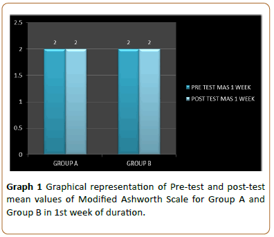
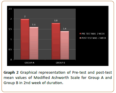
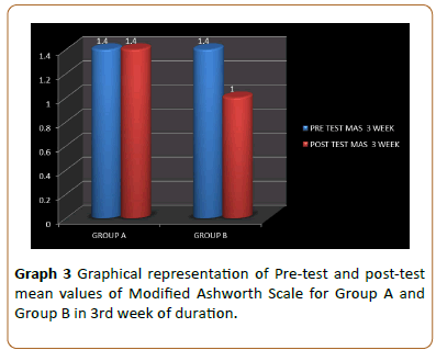
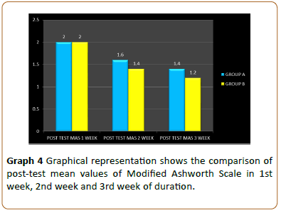
Data analysis was done by IBM SPSS 3620 software. The collected data were analyzed and tabulated with the descriptive and inferential statistics. For the descriptive statistics, the mean and standard deviation were calculated and for the inferential statistics, the parametric variables were treated with t-test. The results were tabulated and the results were plotted accordingly (Tables 1-3; Graphs 1-4).
Results
According to Table 1 the Pre-test and post-test mean value of Modified Ashworth Scale for Group A in 1 week was 2.0000 and pre-test and post-test mean value of Modified Ashworth Scale for Group B in 1 week was 2.0000. No significant difference was found between Conventional group and TENS Group in reducing spasticity (p<0.05).
| Age | Mean | ||||
|---|---|---|---|---|---|
| Gender | |||||
| PRE-TEST MAS GA 1 WEEK | MALE | FEMALE | 2 | 0 | |
| POST-TEST MAS GA 1 WEEK | 2 | 0 | |||
| PRE-TEST MAS GA 2 WEEK | 2 | 0 | |||
| OST TEST MAS GA 2 WEEK | 1.6 | 0.54772 | |||
| PRE-TEST MAS GA 3 WEEK | 45-70 | 4 | 1 | 1.4 | 0.44721 |
| POST-TEST MAS GA 3 WEEK | 1.4 | 0.44721 | |||
| PRE-TEST MAS GB 1 WEEK | 2 | 0 | |||
| POST-TEST MAS GB 1 WEEK | 2 | 0 | |||
| PRE-TEST MAS GB 2 WEEK | 1.8 | 0.44721 | |||
| POST-TEST MAS GB 2 WEEK | 45-70 | 3 | 2 | 1.4 | 0.548 |
| PRE-TEST MAS GB 3 WEEK | 1.4 | 0.548 | |||
| POST-TEST MAS GB 3 WEEK | 1 | 0.548 | |||
Table 1: Pre-test and Post-test mean values of Modified Ashworth Scale for Group A and Group B in 1st week, 2nd week, and 3rd week of duration.
According to Table 1 the Pre-test and post-test mean value of Modified Ashworth Scale for Group A in 2 week was 2.0000 and 1.6000, and the pre-test and post-test mean value of Modified Ashworth Scale for Group B in 2 week was 1.8000 and 1.4000. The Group B showed significantly reduced spasticity after therapeutic intervention than conventional group.
According to Table 1 the pre-test and post-test mean value of Modified Ashworth Scale for Group A in 3 week was 1.4000 and the pre-test and post-test mean value of Modified Ashworth Scale for Group B in 3 week was 1.4000 and 1.0000. The Group B showed significant reduction in spasticity after TENS than conventional group (p<0.05).
According to Table 2 the post-test mean value of Modified Ashworth Scale for Group A was 2.0000 and for Group B was 2.0000 in 1 week. There is no significant difference is found in the reduction of spasticity for both Groups at p<0.05.
| Mean | |
|---|---|
| POST-TEST MAS GA Vs GB 1 WEEK | 2 |
| 2 | |
| POST-TEST MAS GA Vs GB 2 WEEK | 1.6 |
| 1.4 | |
| POST-TEST MAS GA Vs GB 3 WEEK | 1.4 |
| 1.2 |
Table 2: Comparison of post-test means values of Modified Ashworth Scale between Group A and Group B in 1ST week, 2nd week and 3rd week of duration.
According to Table 2 the post-test mean value of Modified Ashworth Scale for Group A was 1.6000 and for Group B was 1.4000 in 2 week. There is statistically significant difference is found in reduction of spasticity for Group B (TENS GROUP) at p<0.05. According to Table 2 the post-test mean value of Modified Ashworth Scale for Group A was 1.4000 and for Group B was 1.2000 In 3 week. There is statistically significant difference is found in reduction of spasticity for Group B (TENS GROUP) at p<0.05.
According to Table 3 the post-test of Modified Ashworth Scale for Group A and Group B in 2 week and post-test of Modified Ashworth Scale for Group A and Group B in 3 week was compared to find out the reduction in spasticity.
| SIG | T | df | Sig 2 tailed | Mean difference | |
|---|---|---|---|---|---|
| POST-TEST MAS GA Vs GB 2 WEEK | 1 | 0.577 | 8 | 0.58 | 0.2 |
| 0.577 | 8 | 0.58 | 0.2 | ||
| POST-TEST MAS GA Vs GB 3 WEEK | 0.252 | 0.632 | 8 | 0.55 | 0.2 |
| 0.632 | 7.692 | 0.55 | 20000 |
Table 3: The table shows the comparison of post-test significant value, t value and mean difference of Modified Ashworth Scale in Group A and Group B in 2ND Week and 3RD Week.
Discussion
To find out the effectiveness of Transcutaneous electrical nerve stimulation TENS) over Gastrocnemius muscle spasticity in stroke patients.
According to the results, Transcutaneous Electrical Nerve Stimulation decreases spasticity effectively when compared to the application of conventional exercise in the 2nd week and in the 3rd week (Table 1). Anti-spastic effects were keenly observed in the conventional group, and it was assumed mainly due to the application of physical therapy during intervention. Similarly, many studies reveal that strokeinduced spasticity are reduced more effectively by transcutaneous electrical nerve stimulation than the exercise alone.
In the present study, electrodes were placed over the bellies of gastrocnemius muscle, which is innervated by the sural nerve, communicating branch of the common peroneal nerve. Other studies reveals that Transcutaneous Electrical Nerve Stimulation, on which the electrodes were applied to acupuncture points or posterior to the fibular head shows the anti-spastic effect and these sites are innervated by common peroneal and sural nerves. Therefore, it is concluded that, though the electrodes were placed on different sites, Transcutaneous Electrical Nerve Stimulation can reduce the spasticity by amplifying presynaptic inhibition on the sural or peroneal nerves.
It has been accepted that the anti-spastic effects of Transcutaneous Electrical Nerve Stimulation, may increases the release of endogenous Gamma Amino Butyric Acid (GABA) and Opiates, by which both act as an inhibitory neurotransmitters, on the dorsal horn of the spinal cord, and this shows the similar achievement on anti-spastic effects as those of baclofen and morphine.
Transcutaneous electrical nerve stimulation (TENS) at two to three times the sensory threshold produces vibrations in stimulated muscles and surrounding regions. Moreover, the rapid stimulation of vibrations triggers primary afferent neurons and increase the release of acetylcholine, a major neurotransmitter in the context of muscle contraction.
However, prolonged stimulation may reduce muscle contraction by lowering the excitability of homonymous motor neurons by depleting acetylcholine, as occurs during muscle fatigue.
At the initial stage, spasticity tends to increase temporarily due to the application of Transcutaneous Electrical Nerve Stimulation but then progressively diminishes at the later stage. Recently, it has been reported that transcutaneous electrical nerve stimulation reduces spasticity and ankle clonus in Upper Motor Neuron lesion by improving the joint movement and gait function.
This study was mainly focused on Gastrocnemius muscle to improve the balance by reducing the muscle spasticity, as it is believed to be the main cause for increasing the proprioception input out of the somatic sense in lower limbs. Transcutaneous Electrical Nerve Stimulation over calf muscle region, which plays a pivotal role in controlling and maintaining the standing posture, probably produces higher somatosensory inputs than the standard rehabilitation.
According to the previous study on the effect of Transcutaneous Electrical Nerve Stimulation on motor cortex excitability, it shows that excitability was deflated in cortical areas corresponding to TENS-stimulated muscles, but elevated in antagonist brain areas.
Stroke patients are commonly associated with ankle plantar flexion contracture due to spasticity of the calf muscle and tibia is anterior weakness. Thus, the present study was mainly focused on the Gastrocnemius muscle to improve the effectiveness in reduction of muscle spasticity.
Ten subjects were selected for this study and they are divided into groups and received the physical therapy as well as the intervention for 3 weeks and by using Modified Ashworth Scale, the spasticity is measured before and after the intervention. The post-test are measured and calculated on the basis of MAS score and their results were tabulated.
However, although a study of Transcutaneous Electrical Nerve Stimulation was found to be reducing spasticity effectively and also reinforcing in the balance among stroke patients, the long term Transcutaneous Electrical Nerve Stimulation application have not been determined. Therefore, more studies can be done to produce the long-term effect on Transcutaneous Electrical Nerve Stimulation on treating the muscle spasticity in stroke patients.
Conclusion
The study concludes that the application of the Transcutaneous Electrical Nerve Stimulation (TENS) can reduce the Gastrocnemius muscle spasticity among stroke patients.
Limitations and recommendations
Limitations:
• The sample size was small, which limits the generalizability of the data.
• There was no long-term follow-up.
• Recommendations:
• Study can be done with larger sample size, for longer duration so it will improve spasticity.
• Study can be done with other muscle group.
Study can be done to know the effect of TENS on gait, cadence, step length, stride length.
References
- P Senthil, Selvam, Arun B (2016) A Study of Neck Pain and Position in Drivers Role of Scapular. J Phys Ther Sci 6: 125-136.
- Kim EK, Kim JS (2016) Correlation between rounded shoulder posture, neck disability indices, and degree of forward head posture. J Phys Ther Sci 28: 2929-2932.
- Mansfield N, Sammonds G, Nguyen (2015) Driver discomfort in vehicle seats e Effect of changing road conditions and seat foam composition. Appl Ergon 2015 50: 153-159.
- smith J, Mansfield N, Gyj N, Pagett M, Bateman B (2012) Driving performance and driver discomfort in an elevated and standard driving position during a driving simulation. J Phys Ther Sci 27: 75-87.
- Noda M, Malhotra R, DeSilva V, Sapukotana P, DeSilva A (2015) Occupational risk factors for low back pain among drivers of three-wheelers in Sri Lanka. Int J Occup Environ Health 21: 216-222.
- Bhavya R (2014) Comparing the effectiveness of movement with mobilization versus mobilization in neck pain among working population. J Phys Ther Sci 25: 865-871.
- Paine R, Voight ML (2013) Invited clinical commentary the role of the scapula. Int J Sports Phys Ther 8: PMC3811730.
- Sinha AK. “Morbidity profile of auto-rickshaw drivers from randomly selected drivers unions in bangalore city”.
- Karthikeyan S (2013) The impact of computer use of neck muscle strength among the enginner college students. J Phys Ther Sci 4: 42-54.
- Suselarayudu (2009) Effect of neck retraction excersise on mechanical neck pain. Cochrane Database Syst Rev 20: CD004250.
- raj M (2009) Efficacy of positional release technique on neck pain due to postural syndrome. J Phys Ther Sci 27: 2461-2464.
- Szeto GP1, Lam P (2007) Work-related Musculoskeletal Disorders in Urban BusDrivers of Hong Kong. J Occup Rehabil 17: 181-198.
- Ariens G, van Mechelen W, Bongers PM, BouterM, Wal G (2000) Physical risk factors for Neck pain. Scand J Work Environ Health 26: 7-19.
- Barr AE, Barbe MF (2004) Inflammation reduces Physiological tissue tolerace in the development of work-related musculoskeletal disorders. J Electromyography kinesiol 14: 77-85.
- Canerio JP, Sullivan p, Burnett A, (2010) The Influence of different sitting postures on head/ Neck posture and muscle activity. J Manual Thera 9: 54-60.
- Giombini A, Di Cesare A, Quaranta F, (2013) Neck balance system in the treatment of chronic Mechanical neck pains: a prospective randomized Control study. Eur J physio Rehabi Medi 8: 283-290.
- Hagberg M (1948) Occupational musculoskeletal Stress on shoulder disorders of the neck and Shoulder: a review of possible pathophysiology. Int Arch Occupatenviron Health 7: 269-278.
- Issever H, Onen L, Sabuncu H, altunkaynak, (2002) Personality characteristics, psychological symptoms and anxiety levels of drivers in charge of urban transportation in Istanbul. Occupational Medicine 52: 297-303.
- Kim TH (2008) The effects of stretching exercise on workers with neck and shoulder pain. Korean J Spor Sci 17: 981-992.
- Kim YM (2009) Effects of the use of the hold relax technique to treat female VDT workers with work related Neck-shoulder complaints. Korean J Occupenviro Med 21: 18 27.
- Kompier MAJ, dimartino V (1995) Review of bus drivers, occupational stress and stress prevention. Stress Medicine 11 253-262
- Lee SY, Lee SC (2011) Mediating effect of coping behavior on the relationship between driving stress and traffic accident risk. Korean Indus Organ Psychol 24: 673-693.
- Ludewig PM, Cook TM, (2000) Alternations in shoulder kinematics and associated muscle activity in people with symptoms of shoulder impingement, world J Sport Sci 3: 276-291.
- Magnusson M, Pope M, Wilder D, Areskoug B (1996) Are occupation drivers at an increased risk for developing musculoskeletal disorders? Physiothera 21: 710-717.
- Ohlsson K, Attewell RG, Palsson B, Karlsson B, balogh I, et al. (1995) repetitive industrial work and neck and upper limb disorders in females. Am J Ind Med 27: 731- 47.
- Pavan EE, Frigo CA, Pedotti A, (2013) Influence Indian Journal of Physiotherapy and Occupational Therapy. Physiothera 10: 951-955.
- Ragland DR, winkleby MA, Schwalbe J, Holman B, Morse L, et al. (1987) Prevalence of hypertension in bus drivers. Int J Epidemiol 16: 208-214.
- Rehn B, Lundström R, Nilsson T, Bergdahl I, Ahlgren C, et al. (2002) Musculoskeletal symptoms among drivers of all-terrain vehicles. J Sound vib 253: 21-29.
- Sluiter J, rest K, Frings-Dresen M (1997) A Critical review of epidemiologic evidence for work Related musculoskeletal disorders of the neck, Upper extremity, lower back. Chapter 2, NIOSH Publication pp: 97-141.
Open Access Journals
- Aquaculture & Veterinary Science
- Chemistry & Chemical Sciences
- Clinical Sciences
- Engineering
- General Science
- Genetics & Molecular Biology
- Health Care & Nursing
- Immunology & Microbiology
- Materials Science
- Mathematics & Physics
- Medical Sciences
- Neurology & Psychiatry
- Oncology & Cancer Science
- Pharmaceutical Sciences
