Effect of Knee-Ankle-Foot Orthosis (KAFO) on Knee Kinematics and Kinetics in an Individual with Knee Varus Alignment
Huda Hamdan AL-Fatafta*
Orthotics and Prosthetics Department, University of Jordan, Jordan
- *Corresponding Author:
- Huda Hamdan AL-Fatafta
Orthotics and Prosthetics department
university of Jordan, Jordan
Tel: 00962796497364
E-mail: huda_alfatafta@hotmail.com
Received date: November 08, 2016; Accepted date: November 26, 2016; Published date: November 30, 2016
Citation: Huda Hamdan AL-Fatafta (2016) Effect of Knee-Ankle-Foot orthosis (KAFO) on knee kinematics and kinetics in an individual with knee varus alignment. Pro Ort Open J 1:2.
Copyright: © 2016 Fatafta HHA. This is an open-access article distributed under the terms of the Creative Commons Attribution License, which permits unrestricted use, distribution, and reproduction in any medium, provided the original author and source are credited.
Short Commentary
The medial compartment knee osteo arthritis (OA) is the most affected location for OA [1]. The main knee biomechanics changes which are associated with the medial compartment knee OA are less knee flexion value [2-4] (the normal mean maximum knee flexion has been shown to be 64.6° during walking, 98.6° during ascending, and 90.3 °during descending) [5], high knee varus angle and an increased the external knee adduction moment (EKAM) during walking and stairs climbing due to shifting the knee joint centre more laterally and the centre of the load medially [6]. These changes lead to pain during daily activities and progression of the knee OA [7,8]. The EKAM is considered as the most important variable in the frontal plan and has two peaks: The first peak is a sharp one after initial contact during early stance phase, while the second one is in the late stance phase. The first peak is affected by the amount of knee varus, joint space narrowing, OA severity, and progression level, while the second one is more correlated to the amount of toe out and pain levels [9].
Various of orthotics treatments have been used to reduce the EKAM and knee varus degree and to increase knee flexion degree. The custom made and off the shelf (OTS) knee valgus braces are one of current orthotics treatments. However, some investigations have found no significant reductions in the EKAM during walking [10-12] or during stair climbing [12] especially with severe knee varus degree. In addition, walking with knee flexion reduction during swing phase has been found [13].
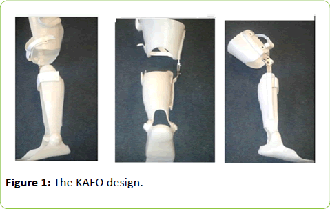
A custom made knee ankle foot orthosis (KAFO) with three point pressure correction was one of a suggested alternative treatment to reduce the EKAM and increase knee flexion value during walking and stairs climbing for severe knee varus participants. The KAFO design includes: a single upright, the Ottobock knee joint (17lk1=L/R1-5), with a 5° knee flexion stop, also with a simple hinged ankle joint to allow free movement and manufactured in 4.5 mm copolymer polypropylene (Figure 1).
One male individual with a 10° knee varus deformity (aged 45, mass 85 kg, height 1.68 m) participated in this study without any previous knee injuries. The data were collected in the gait laboratory using 16 infra red Qualisys OQUS cameras, (Qualisys AB, Gothenburg, Sweden), and 2 embedded AMTI force plates in a walkway (AMTI: Advanced Mechanical Technology Incorporation, Watertown, USA, model-BP600400).
The result shows that the KAFO significantly reduced the knee varus angle compared to the shoe and both knee valgus braces during walking and stair climbing with decreases of up to 12 degrees. Moreover, the KAFO reduced the first peak of the EKAM by a greater margin than either of the knee valgus braces (11.4%, 15%, and 12.6% compared to the shoe, OTS and custom knee valgus braces, respectively) during walking. The KAFO increased the knee flexion angle at initial contact (IC) significantly compared to the shoe and OTS during walking (mean difference 8.6, 4.1degrees respectively). However, no significant changes were seen during stair climbing, and this could be due to a high knee varus angle which was seen (up to 30 degree) and the interventions cannot reduce it efficiently (Tables 1 and 2).
| Variables | Mean ± SD | P value | |||||||||
|---|---|---|---|---|---|---|---|---|---|---|---|
| EKAM first Peak | Shoe | OTS | Custom | KAFO | Shoe vs. OTS | Shoe vs. Custom | Shoe vs. KAFO | KAFO vs. Custom | KAFO vs. OTS | Custom vs. OTS | |
| Wa | 0.70 ± 0.01 | 0.71 ± 0.01 | 0.73 ± 0.02 | 0.62 ± 0.0 | 1.0 | 1.0 | 0.04 | 0.07 | 0.07 | 1.0 | |
| As | 0.85 ± 0.3 | 0.79 ± 0.4 | 0.78 ± 0.02 | 0.79 ± 0.05 | 1.0 | 1.0 | 1.0 | 1.0 | 1.0 | 1.0 | |
| EKAM second peak | De | 0.81 ± 0.1 | 0.90 ± 0.2 | 0.97 ± 0.6 | 0.81 ± 0.4 | 0.17 | 0.38 | 1.0 | 0.86 | 0.46 | 1.0 |
| Wa | 0.65 ± 0.01 | 0.63 ± 0.02 | 0.65 ± 0.01 | 0.57 ± 0.01 | 1.0 | 1.0 | 0.09 | 0.0 | 0.07 | 1.0 | |
| As | 0.46 ± 0.01 | 0.48 ± 0.02 | 0.45 ± 0.02 | 0.41 ± 0.02 | 1.0 | 1.0 | 0.78 | 0.05 | 0.17 | 1.0 | |
| Knee flexion moment | De | 0.75 ± 0.02 | 0.75 ± 0.01 | 0.77 ± 0.02 | 0.74 ± 0.01 | 1.0 | 1.0 | 1.0 | 1.0 | 1.00 | 1.0 |
| Wa | 0.48 ± 0.4 | 0.55 ± 0.03 | 0.58 ± 0.02 | 0.70 ± 0.04 | 1.00 | 0.27 | 0.33 | 0.60 | 0.42 | 1.00 | |
| As | 0.80 ± 0.5 | 0.82 ± 0.02 | 0.78 ± 0.03 | 0.82 ± 0.03 | 1.00 | 1.00 | 1.00 | 1.00 | 1.00 | 1.00 | |
| De | 1.00 ± 0.03 | 0.98 ± 0.02 | 0.98 ± 0.02 | 1.03 ± 0.03 | 1.00 | 1.00 | 1.00 | 1.00 | 1.00 | 1.00 | |
Table 1: Mean and standard deviation and p value of the knee adduction moment and knee flexion and extension moment among different conditions. Bold result shows significant result run with (ANOVA) with a post-hoc bonferroni correction. Wa: walking, As: ascending, De: descending. Bold indicates significance.
| Variables | Mean ± SD | P value (CI 95%) | |||||||||
|---|---|---|---|---|---|---|---|---|---|---|---|
| Knee flexion at IC | Shoe | OTS | Custom | KAFO | Shoe vs. OTS | Shoe vs. Custom | Shoe vs. KAFO | KAFO vs. Custom | KAFO vs. OTS | Custom vs. OTS | |
| Wa | 2.5 ± 0.6 | 7.0 ± 0.5 | 7.8 ± 0.6 | 11.1 ± 0.6 | 0.03 | 0.02 | 0.00 | 0.28 | 0.03 | 1.0 | |
| As | 66.8 ± 1.1 | 65.2 ± 0.7 | 67.2 ± 0.9 | 67.3 ± 0.6 | 1.0 | 1.0 | 1.0 | 1.00 | 0.36 | 1.0 | |
| Knee flexion at mid swing | De | 14.7 ± 0.4 | 19.9 ± 0.4 | 17.9 ± 0.5 | 15.6 ± 0.8 | 0.01 | 0.10 | 1.00 | 0.31 | 0.01 | 0.00 |
| Wa | 74.1 ± 0.2 | 70.5 ± 3.2 | 72.3 ± 1.3 | 72.4 ± 0.4 | 1.0 | 1.0 | 0.31 | 1.0 | 1.0 | 1.0 | |
| As | 107.0 ± 0.8 | 103.1 ± 0.8 | 100.3 ± 1.5 | 101.5 ± 1.7 | 0.01 | 0.24 | 0.05 | 1.0 | 1.0 | 1.0 | |
| Maximum knee adduction angle | De | 105.9 ± 1.4 | 104.3 ± 1.2 | 102.9 ± 1.2 | 98.8 ± 0.8 | 1.0 | 1.0 | 0.15 | 0.05 | 0.22 | 1.0 |
| Wa | 17.4 ± 0.4 | 11.9 ± 0.3 | 12.2 ± 0.1 | 9.20 ± 0.2 | 0.00 | 0.00 | 0.00 | 0.00 | 0.04 | 1.0 | |
| As | 30.3 ± 0.6 | 30.5 ± 0.3 | 33.5 ± 0.3 | 18.3 ± 0.1 | 1.0 | 0.08 | 0.00 | 0.00 | 0.00 | 0.00 | |
| De | 21.4 ± 0.5 | 19.9 ± 0.4 | 22.2 ± 0.4 | 15.4 ± 0.3 | 0.03 | 1.0 | 0.00 | 0.00 | 0.01 | 0.10 | |
Table 2: Mean and standard deviation (SD) of knee angle in sagittal and frontal planes among conditions. Bold results show significant result according to (ANOVA) was run with 4 factors (shoe, off-the-shelf (OTS), custom knee brace, and KAFO) with a post-hoc bonferroni correction. Wa: walking, As: ascending, De: descending. IC: initial contact.
The efficiency of a KAFO in reducing a knee varus angle is mainly related to the offset joint which was used to correct knee deformity in the frontal plane (knee valgus/varus), with the length of the KAFO applying more force over a more extensive tissue area than knee valgus braces. This meant that it would theoretically be able to correct the deformity more effectively by shift the ground reaction force (GRF) more laterally and the knee joint centre more medially, thereby reducing the EKAM.
Moreover, the KAFO has a more intimate and extensive fit on the lower limb than that provided by an OTS device which is able to keep the tibia and foot in a corrected position via an ankle foot orthosis (AFO) section. This would encourage a reduction in knee varus deformity, prevent hyperextension, and potentially improve knee flexion by shifting the bodies load anterior to the hip and posterior to the knee joint [14].
Additional work will be needed to further evaluate the clinical and biomechanical benefits of a KAFO in a larger number of subjects with different severities of uni-compartmental knee OA. Also, evaluate the long term using benefits is required.
References
- Buckwalter JA, Saltzman C, Brown T (2004) The impact of osteoarthritis: implications for research. Clin Orthop Relat Res : S6-15.
- Al-Zahrani KS, Bakheit AM (2002) A study of the gait characteristics of patients with chronic osteoarthritis of the knee. Disabil Rehabil 24: 275-280.
- Mündermann A, Asay JL, Mündermann L, Andriacchi TP (2008) Implications of increased medio-lateral trunk sway for ambulatory mechanics. J Biomech 41: 165-170.
- Walker CR, Myles C, Nutton R, Rowe P (2001) Movement of the knee in osteoarthritis. The use of electrogoniometry to assess function. J Bone Joint Surg Br 83: 195-198.
- Jevsevar DS, Riley PO, Hodge WA, Krebs DE (1993) Knee kinematics and kinetics during locomotor activities of daily living in subjects with knee arthroplasty and in healthy control subjects. Phys Ther73: 229-239.
- Hicks-Little C A, Peindl RD, Hubbard TJ, Scannell BP, Springer BD, et al. (2011) Lower extremity joint kinematics during stair climbing in knee osteoarthritis. Med Sci Sports Exerc43: 516-524.
- Bennell KL, Bowles KA, Wang Y, Cicuttini F, Davies-Tuck M, et al. (2011) Higher dynamic medial knee load predicts greater cartilage loss over 12 months in medial knee osteoarthritis. Ann Rheum Dis70: 1770-1774.
- Miyazaki T, Wada M, Kawahara H, Sato M, Baba H, et al. (2002) Dynamic load at baseline can predict radiographic disease progression in medial compartment knee osteoarthritis. Ann Rheum Dis61: 617-622.
- Sasaki K, Neptune RR (2010) Individual muscle contributions to the axial knee joint contact force during normal walking. J Biomech43: 2780-2784.
- Pollo FE, Otis JC, Backus SI, Warren RF, Wickiewicz TL (2002) Reduction of medial compartment loads with valgus bracing of the osteoarthritic knee. AmJSports Med 30:414-421.
- Gaasbeek RD, Groen BE, Hampsink B, van Heerwaarden RJ, Duysens J (2007) Valgus bracing in patients with medial compartment osteoarthritis of the knee. A gait analysis study of a new brace. Gait Posture 26: 3-10.
- 12. Al-Zahrani Y (2014) Effectiveness of a valgus knee brace on biomechanical and clinical outcomes during walking and stair climbing in individuals with knee osteoarthritis. PhD thesis. University of Salford. Available at: https://usir.salford.ac.uk/32833/1/Yousef_Al-zahrani_Thesis_%28Sep-2014%29.pdf. Accessed April 5, 2016.
- Jones RK, Nester CJ, Richards JD, Kim WY, Johnson DS, et al. (2013) A comparison of the biomechanical effects of valgus knee braces and lateral wedged insoles in patients with knee osteoarthritis. Gait Posture 37: 368-372.
- Johnson GR, Ferrarin M, Harrington M, Hermens H, Jonkers I, et al. (2004) Performance specification for lower limb orthotic devices.Clin Biomech 19: 711-718.

Open Access Journals
- Aquaculture & Veterinary Science
- Chemistry & Chemical Sciences
- Clinical Sciences
- Engineering
- General Science
- Genetics & Molecular Biology
- Health Care & Nursing
- Immunology & Microbiology
- Materials Science
- Mathematics & Physics
- Medical Sciences
- Neurology & Psychiatry
- Oncology & Cancer Science
- Pharmaceutical Sciences

