Diaphragmatic Hernia and Chest Pain
Criado-Álvarez JJ1-3* and González J1,3
1Department of Occupational Therapy, Speech Therapy and Nursing, University of Castilla-La Mancha, Calle Altagracia, Real, Spain
2Department of Physical Activity and Sport Sciences, European University of Madrid, Calle Tajo, Villaviciosa de Odón, Madrid, Spain
3Health Service of Castilla-La Mancha, Calle Mateo Ramos de la Nava, San Bartolomé de las Abiertas, Toledo, Spain
- *Corresponding Author:
- Criado-Álvarez JJ
Health Service of Castilla-La Mancha (SESCAM)
Calle Mateo Ramos de la Nava
7 45654 San Bartolomé of the Abiertas
Toledo, Spain
Tel: +34925704020
E-mail: jjcriado@sescam.jccm.es
Received Date: December 10, 2016; Accepted Date: December 27, 2016; Published Date: January 03, 2017
Citation: Criado-Álvarez JJ, González J. Diaphragmatic Hernia and Chest Pain. J Clin Med Ther. 2017, 2:1.
Copyright: © 2017 Criado-Álv arez JJ , et al. This is an open-accessa rticle distributed under the terms of the Creative Commons Attribution License, which permits unrestricted use, distribution and reproduction in any medium, provided the original author and source are credited.
Abstract
It’s a case of chest pain in a patient with a history of ischemic heart disease. She present in emergency room an increase of pain with respiratory movement, conscious and good general condition. The chest X-ray has a suggestive image of hiatus hernia with a herniation of the stomach, is confirmed in the CT scan. Gastroscopy confirmed the diagnosis, and aspirated the gas from stomach. The patient remains asymptomatic, and without pain at 24 hours and at 6 months control.
Keywords
Congestive heart failure; Ischemic heart disease; Diaphragmatic Hernia
Diaphragmatic Hernia and Chest Pain
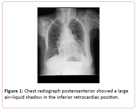
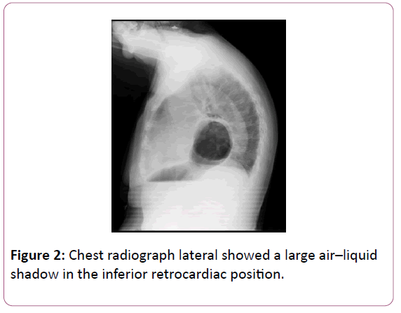
This is a 94-year-old patient with a history of ischemic heart disease and congestive heart failure, who is referred by her family doctor to the hospital for an epigastric pain with a 15- day chest irradiation [1,2]. The pain increases with inspiration and partially yields with paracetamol 650 mg orally. No cough, dyspnoea, orthopnea, heartburn, nausea or vomiting, or abdominal discomfort. Upon arrival to the emergency room, the patient is conscious, with good general condition and perfusion, resting and hemodynamically stable eupneic [3-5]. Clinical examination has arrhythmic cardiac sounds, no murmurs, and slight decrease in murmur vesicular. On the chest X-ray, there is a suggestive image of hiatus hernia (Figures 1 and 2).
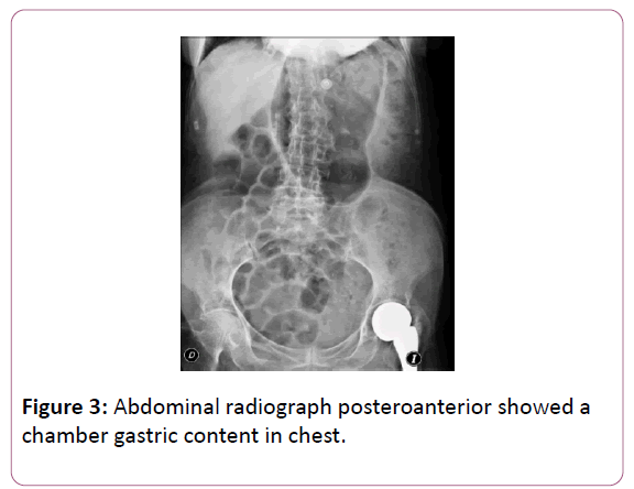
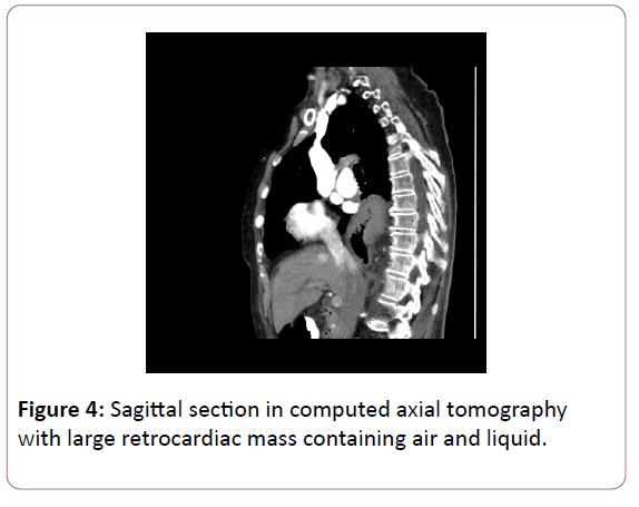
On abdominal radiography a herniation of the stomach with rotation to the thoracic cavity (Figure 3), this is confirmed in the CT scan (Figure 4) [6-7].
These hernias are usually congenital in origin and can increase in size over time. Gastroscopy is performed confirming the diagnosis, allowing the aspiration of gas from the gastric chamber. The degree of displacement can produce obstructive symptoms of chest pain as occurred in this patient. The patient remains asymptomatic, and without pain so is discharged at 24 hrs. In chest X-ray of control at 6 months, remains part of the hernia and the patient remains stable and asymptomatic [8-10].
References
- Yasin M, Reed JC (2016) Diaphragmatic Hernia causing lung collapse. N Engl J Med 374: 1874.
- Malekzadegan A, Sargazi A (2016) Congenital Diaphragmatic Hernia with Delayed Presentation. Case Rep Surg 2016: 4.
- Kohli N, Mitreski G, Yap CH, Leong M (2016) Massive symptomatic right-sided Bochdalek hernia in an adult man. BMJ Case Rep.
- Manson HJ, Goh YM, Goldsmith P, Scott P, Turner P (2016) Congenital diaphragmatic hernia causing cardiac arrest in a 30-year-old woman. Ann R Coll Surg Engl, pp: e1-e3.
- Pattnaik MK, Sahoo SP, Panigrahy SK, Nayak KB (2016) Morgagni hernia: A rare case report and review of literature. Lung India 33: 427-429.
- Sousa CC, Duarte J (2016) Large Hiatal Hernia. N Engl J Med 375: 2081.
- Palios J, Clements S, Lerakis S (2014) Chest pain due to hiatal hernia mimicking as cardiac mass. Acute Card Care 16: 88-89.
- Lloret CB, Damiá AB, Morón CC, Montaño JC (2015) Incidental finding of diaphragmatic hernia during 99mTc-tetrofosmin photon emission computed tomography. Med Clin 144: 481.
- Deressa BK, Bruyninx L, Ngassa M, Thill V, Toussaint E (2016) Uncommon cause of retrosternal pain. Acta Gastroenterol Belg 79: 251-253.
- Ghosh RK, Fatima K, Ravakhah K, Hassan C (2016) Gastric volvulus: an easily missed diagnosis of chest pain in the emergency room. BMJ Case Rep.
Open Access Journals
- Aquaculture & Veterinary Science
- Chemistry & Chemical Sciences
- Clinical Sciences
- Engineering
- General Science
- Genetics & Molecular Biology
- Health Care & Nursing
- Immunology & Microbiology
- Materials Science
- Mathematics & Physics
- Medical Sciences
- Neurology & Psychiatry
- Oncology & Cancer Science
- Pharmaceutical Sciences
