Coronary Artery Disease (CAD): Correlation between Predisposing Factors and the Affected Coronary Arterial Branches
Mo’ath S. Alzu’bi*
Department of Basic Medical Science, University of Hashemite, Jordan
- *Corresponding Author:
- Mo’ath S. Alzu’bi,
Department of Basic Medical Science,
Hashemite University,
Jordan,
Tel: 00962797443240;
E-mail: moath@hu.edu.jo
Received Date: May 28, 2021; Accepted Date: June 11, 2021; Published Date: June 18, 2021
Citation: Alzu’bi MS (2021) Coronary Artery Disease (CAD): Correlation between Predisposing Factors and the Affected Coronary Arterial Branches. J Anat Sci Res Vofl. 4 No.4:3.
Abstract
It is well-established that coronary artery disease (CAD) is the leading cause of death worldwide. CAD is caused by an imbalance between blood perfusion to the myocardium and its nutritional demands as a result of atherosclerotic obstruction of the coronary arteries. Several modifiable and non-modifiable risk factors have been blamed to cause CAD. The literature contains huge amount of studies about CAD and its risk factors but very little information is present in the literature concerning the effect of different risk factors on specific coronary arteries or their respective branches. Taking into account the lifestyle of the Jordanian population, the contribution of each risk factor as a causative factor for CAD and the possible linkage between different risk factors and the coronary branches affected by CAD have never been studied. In this study, six coronary arterial branches were included. These branches are the right coronary artery (RCA), the left main (LM) coronary artery, the left circumflex (LCx), the left anterior descending (LAD) artery, the obtuse marginal (OM) artery and the diagonal (Diag.) artery. Data From 600 CAD patients attending the Cardiology clinic at King Abdullah University Hospital (KAUH) were included. The collected data include detailed history of the predisposing CAD risk factors and the diseased coronary branches were recorded for each patient. Patients data was analyzed by Chi-Square tests using SPSS software and p=0.05 was considered as significant.81.2% of the patients were males and 75.2% of the patients occur in the age range between (41-64) years old which are, in this thesis, arbitrarily referred to as middle-aged patients. This study found some interesting results concerning the possible link between CAD-affected coronary branches and the underlying risk factors.
Keywords
Coronary arteries; Ischemic heart; Coronary artery predilection; Hypertension
Introduction
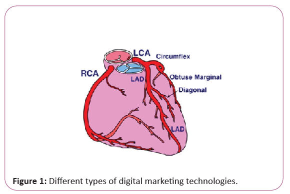
Narrowing of coronary arteries due to atherosclerosis can affect any of the coronary arteries, singly or in any combination. Clinically significant atherosclerotic plaques can be located anywhere but tend to develop within the first several centimeters of the LAD and LCx, and along the entire length of the RCA [1] (Figure 1).
It is well-known that the risk factors for CAD can be categorized into two groups; modifiable and non- modifiable (or constitutional) risk factors. The modifiable risk factors are acquired or related to modifiable behavior whereas the constitutional risk factors are less controllable. Table 1 illustrates the factors of concern into these two groups
| Modifiable risk factors | Non-modifiable risk factors |
|---|---|
| Diabetes Mellitus (DM) Hypertension (HTN) Dyslipidemia |
Family history Increasing age Male sex |
Table 1: The modifiable and non-modifiable risk factors for CAD.
When reviewing the literature, there seems to be some connections between the predisposing factors and the coronary artery branches affected by CAD. Starting with modifiable risk factors with regards to DM, a study found that the coronary vessels affected are ordered from the most to the least common as follows; the RCA, then the LCx artery, then the LAD artery, and lastly the LM artery [2]. Another study have shown different results for coronary branches affected in diabetics, that are; the most commonly involved coronary artery in diabetic patients was the LCx, followed by the LAD, and the RCA and LM arteries, while, in non-diabetic patients, the most commonly diseased coronary artery was LAD, followed by the LCx, and the RCA and LM arteries [3].
When CAD is present in HTN patients, it is significantly more frequently to be localized in the LAD artery [4]. Regarding dyslipidemia patients, it has been shown that the most significant frequently diseased coronary artery is the LAD artery, followed by the LCx and the RCA. But no significant association is found with the LM coronary artery [5]. Regarding smoker CAD patients, the most significant association was found with the RCA followed by the LCx then the LAD artery [6].
Moving to the non modifiable risk factors, a study performed on young CAD patients, under the age of 40 years have shown that the most common location of significant atherosclerotic coronary lesions was the LAD artery (61.6%) followed by the RCA (27.4%) [7]. Another study performed on CAD patients younger than 35 years old have shown that almost 50% of the lesions occurred in LAD followed by 25% in RCA and 20% in LCx artery, while only one patient (1%) had isolated significant CAD of LM coronary artery [8].
In females of all age groups, the pattern of coronary arteries involved in disease is the same. That is the LAD is the most commonly CAD-involved artery, followed by LCx and RCA and LM artery is the least commonly involved artery [9].
Stenosis of the LM artery is a relatively uncommon but it is a significant cause of increased morbidity and mortality among patients with CAD [9].
After reviewing the literature, it seems that there are no previous studies that link different CAD-affected coronary branches with the underlying predisposing factors. It is of great importance to try to find the possible link between the affected coronary arteries and the underlying risk factors as it might help the diagnosis and treatment planning of CAD. It is the primary goal of this study to navigate for the possible link that might exist between the affected coronary arteries and the predisposing risk factors.
Materials and Methods
Relevant data from 600 CAD patients were collected retrospectively in a 6-months time interval between February 2018 and August 2018 from King Abdullah University Hospital (KAUH) in cooperation with cardiac surgery and cardiology departments.
All of the patients underwent catheterization before. For each patient, the previous visits, discharge summaries, lab tests (for lipid profile) and the treatment done were studied to get information about clinical and angiographic profiles of CAD patients.
Non-modifiable risk factors were recorded (age, sex, and family history). Also the presence of modifiable risk factors were recorded (HTN, DM, smoking and lipid profile). In this study, positive family history is defined as the presence of CAD in the first degree relatives (siblings and offsprings).
Regarding the lipid profile, the exact values were taken for LDL, HDL, TG, and TC in (mmol/liter) before they were catheterized according to KAUH reference values (Appendix A).
In this study the significantly diseased coronary artery is defined as one or more of the following:
1- Occlusion is equal to or greater than 50% of the caliber of artery.
2- The artery has been operated before (i.e. stented, ballooned or bypass-grafted).
The mean age of the sample was 56.50 ± 10.18. 487 patients were males, 399 patients were smokers, 420 patients were hypertensive, 320 patients were diabetic, 301 patients had positive family history and 471 patients were dyslipidemic. Of the 600-patient sample, 18 patients underwent bypass grafting on previously stented arteries. Two types of by-pass grafting were included as they are the modes of surgery that were done for some of the patients: the saphenous vein grafts (SVG) and the left internal mammary artery (LIMA) grafts.
Six coronary branches were studied in our analysis which is namely, the:
1- Right Coronary Artery (RCA)
2- Left Main Coronary Artery (LM)
3- Left Circumflex Artery (LCx)
4- Left Anterior Descending Artery (LAD)
5- Obtuse Marginal Artery (OM)
6- Diagonal Artery (Diag)
When defining the presence of risk factors; smoking, HTN, DM and family history were defined as present or not, regardless of the amount and duration of smoking, the exact values of blood pressure, the exact sugar levels and the number of first degree relatives affected by CAD. It's obvious that the such details (e.g. amount and duration of smoking) might be important but these details were unavailable and these details are beyond the scope of this study which is the first of a kind to be done, especially for the Jordanian population. These details might be the subject for further studies to promote or negate the initial findings of this study.
According to the patient's age, three age groups were defined. The young age group comprises patients up to the age of 40 years. The middle age group comprises patients who are between 41-64 years. And the old age group comprises patients who are 65 years or older. This age classification is different from that provided by the World Health Organization (WHO). It is based on the risk of CAD among age groups that we defined..
Patients' data were recorded using Microsoft Excel 2010 software as an excel spreadsheet. The relevant patients' data were analyzed using the SPSS version 20 software. The exact level of significance were calculated based on performing Chi-Square tests.
Results
The following two tables show the distribution of the coronary branches affected by disease in CAD patients with regard to the presence or absence of modifiable and non-modifiable risk factors respectively. Below each table, the statistically significant connections between the predisposing risk factors and the coronary branches affected by CAD in Tables 2-4 are highlighted. Coronary artery disease (CAD) remains the leading cause of mortality and morbidity around the world. It is well-known that several classical risk factors are associated with CAD. The modifiable risk factors are acquired or related to the lifestyle and hence, they are subject to modification or can be more controlled. These include diabetes mellitus (DM), hypertension (HTN), dyslipidemia and cigarette smoking. The non-modifiable risk factors are less controllable and include positive CAD family history as genetic polymorphisms, age and sex [1]. Both groups of risk factors do not occur singularly but rather as combinations of two or more factors. Thus identifying the contribution of those individual risk factors to CAD is difficult and has not been addressed in literature.
|
LM |
LAD |
LCx |
RCA |
OM |
Diag. |
|---|---|---|---|---|---|---|
| DM/CAD | p-value=0.47 | p-value=0.12 | p-value=0.10 | p-value=0.001 | p-value<0.0001 | p-value=0.35 |
| +/+ | 10 | 257 | 166 | 216 | 81 | 54 |
| +/- | 310 | 63 | 154 | 104 | 239 | 266 |
| -/+ | 10 | 213 | 130 | 154 | 43 | 13 |
| -/- | 270 | 67 | 150 | 126 | 237 | 100 |
| HTN/CAD | p-value=0.41 | p-value=0.004 | p-value=0.41 | p-value=0.03 | p-value=0.54 | p-value=0.13 |
| +/+ | 15 | 342 | 209 | 270 | 82 | 73 |
| +/- | 405 | 78 | 211 | 150 | 338 | 347 |
| -/+ | 5 | 128 | 87 | 100 | 35 | 24 |
| -/- | 175 | 52 | 93 | 80 | 145 | 156 |
| DL/CAD | p-value=0.01 | p-value=0.09 | p-value=0.27 | p-value=0.34 | p-value=0.46 | p-value=0.07 |
| +/+ | 11 | 363 | 236 | 293 | 91 | 82 |
| +/- | 460 | 108 | 235 | 178 | 380 | 389 |
| -/+ | 9 | 107 | 60 | 77 | 26 | 15 |
| -/- | 120 | 22 | 69 | 52 | 103 | 114 |
| Smok./CAD | p-value=0.34 | p-value=0.10 | p-value=0.48 | p-value=0.13 | p-value=0.31 | p-value=0.41 |
| +/+ | 12 | 302 | 196 | 153 | 75 | 66 |
| +/- | 387 | 97 | 203 | 146 | 324 | 333 |
| -/+ | 8 | 168 | 100 | 117 | 42 | 31 |
| -/- | 193 | 33 | 101 | 84 | 159 | 170 |
Table 2: Distribution of the coronary branches affected by disease in patients with modifiable risk factors, Table 2 suggests the following connections: 1 Diabetes mellitus predispose to the OM artery disease and RCA disease. 2 Hypertension predispose to the LAD artery disease and RCA disease. 3 Dyslipidemia predispose to LM artery disease. 4 Smoking appears to have no significant predisposition to specific coronary artery disease.
|
LM |
LAD |
LCx |
RCA |
OM |
Diag. |
|---|---|---|---|---|---|---|
| Fam.Hx/CAD | p-value=0.03 | p-value=0.43 | p-value=0.47 | p-value=0.48 | p-value=0.06 | p-value=0.53 |
| +/+ | 14 | 238 | 150 | 186 | 67 | 49 |
| +/- | 287 | 63 | 151 | 115 | 234 | 252 |
| -/+ | 5 | 232 | 146 | 182 | 50 | 48 |
| -/- | 292 | 65 | 151 | 115 | 247 | 249 |
| Sex(M)/CAD | p-value=0.01 | p-value=0.32 | p-value=0.22 | p-value=0.04 | p-value=0.18 | p-value=0.09 |
| +/+ | 20 | 379 | 236 | 309 | 99 | 84 |
| +/- | 467 | 108 | 251 | 178 | 388 | 403 |
| -/+ | 0 | 91 | 60 | 61 | 18 | 13 |
| -/- | 113 | 22 | 53 | 52 | 95 | 100 |
| Age/CAD | p-value=0.17 | p-value=0.55 | p-value<0.0001 | p-value=0.02 | p-value=0.41 | p-value=0.37 |
| Young /+ | 0 | 21 | 12 | 15 | 7 | 7 |
| Young /- | 28 | 7 | 16 | 13 | 21 | 21 |
| Middle /+ | 13 | 350 | 205 | 267 | 91 | 73 |
| Middle /- | 438 | 101 | 246 | 184 | 360 | 378 |
| Old / + | 7 | 99 | 79 | 88 | 19 | 17 |
| Old / - | 114 | 22 | 42 | 33 | 102 | 104 |
Table 3: distribution of the coronary branches affected by disease in patients with non modifiable risk factors. Table 3 suggests the following connections: 1 Positive family history predispose to the LM artery disease. 2 Male sex predispose to the LM artery and RCA disease. 3 Middle age predispose to the LCx artery disease and RCA disease
| Coronary artery branch | Associated risk factors (p-value) |
|---|---|
| LM | Dyslipidemia (0.01), Male Sex (0.01), Positive Family History (0.03) |
LAD |
Hypertension (0.004) |
LCx |
Middle Age (<0.0001) |
RCA |
DM (0.001), Middle Age (0.02), Hypertension (0.03), Male Sex (0.04) |
OM |
DM (<0.0001) |
Diagonal |
No Significant Association |
Table 4: Correlations between Predisposing Factors and The Affected Coronary Arterial Branches. To sum up the statistically significant connections in table 2 and table 3, the following Table 4 illustrates the associated risk factors for each of the coronary branches affected by disease from the strongest to the weakest.
Very few articles have reported the frequency, variation of coronary arteries affected and gender differences of CAD. As far as we know, none of the reported publications investigated the link between different risk factors and specific coronary artery affected and this promoted us to undertake this study.
Discussion
The primary objective for this study is to try to find the possible correlations between different risk factors present in CAD patients and the CAD-affected coronary arterial branches. Some interesting associations seem to be present between the two. Our results are based on provisional data analysis of multi-variables using Chi- Square system.
The following paragraphs discuss the possible correlations that might exist between the coronary branches affected by disease and the underlying predisposing factors comparing our results to the scarce amount of information that are found in somewhat similar previous studies in the literature.
CAD and modifiable risk factors
Our results indicate that the most vulnerable coronary branches in DM patients are the OM artery followed by the RCA. Noting that the OM artery is not frequently studied, these results are consistent with findings that shows that the most commonly CAD-affected artery in DM patients is the RCA followed by the LCx artery [2].
Our results indicate that the most vulnerable coronary branches in HTN patients are the LAD artery followed by the RCA. The former finding is consistent with findings that show that the LAD artery is significantly more involved in CAD in the hypertensive patient [4].
Our results indicate that the most vulnerable coronary branch in dyslipidemia patients is the LM coronary artery. This opposes findings that claim that there is no significant association is present between dyslipidemia and LM disease. This study found that, among dyslipidemia patients, the most significant association was with LAD, followed by LCx and RCA [5].
Our results indicate there is no specific association between smoking and the coronary branches affected by disease. This is inconsistent with findings that shows that the most significant association in smokers is found with the RCA, followed by the LCx and the LAD arteries [6].
Cad and non-modifiable risk factors
Our results indicate that the most vulnerable coronary branch in CAD-positive family history patient is the LM coronary. As far as we know, no relevant information is present in the literature regarding the linkage between CAD-positive family history and the affected coronary branches.
In our sample 81.7% were males. This percentage is approximately the same as the percentage found by which is 81.73% [8]. and very near to the percentage found by which is 86.2% [10]. It's worth mentioning that the aforementioned percentages of the related studies concerned the young CAD patients (less than 40 years) which appear to hold true for CAD patients in general.
Our results indicate that the most vulnerable coronary branches in males are the LM followed by the RCA. As far as we know, no relevant information is present in the literature regarding the linkage between sex and the affected coronary branches.
Regarding age, our results indicate that of the three age groups the middle age group (41-64 years) are mostly affected which made up (75.2%) of the sample[11]. This was followed by the old group (65 years or more) which made up (20.2%) of the sample and lastly the young group (below 40 years) was the least affected which made up (4.7 %) of the total sample. These results are in accordance with claims that the risk of CAD increases five-fold between the 40-60 years of age which is around the age range that we defined as middle age [1].
Our results indicate that the most vulnerable coronary branch in the middle age group is LCx artery followed by the RCA. As far as we know, no relevant information is present in the literature regarding the linkage between age and the affected coronary branches.
Our results indicate some sort of association between the coronary branches affected by CAD and the underlying predisposing factors. This is of a great clinical importance as it would aid in the diagnosis and treatment planning for different risk factors affecting CAD patients. The exact mechanism by which different predisposing factors tend to affect different coronary branches is not known but it could be related to different dimensions (length and diameter) of the different coronary arteries and the possible different metabolic needs for different parts of the heart myocardium supplied by respective branches of coronary arteries. Gender differences could be attributed to the different habits present between the two sexes (e.g. males smoke more) and to the different hormonal status present between males and females. Further future studies are required to support the finding of our study.
Conclusion
Although the arterial structure is similar and the histo-pathological changes of atherosclerosis are the same in all arteries, the exact reason why different predisposing factors selectively affect different coronary arteries is not known. Furthermore, since there are no previous reports in the literature explaining that, we presume that due to different metabolic needs of different parts of the myocardium and unknown patho-physiological players to cause CAD. Further patho-physiological studies are needed to illustrate that selective mechanism.
References
- Vinay Kumar, Abul K.Abbas, John C.Aster (2013) Robins Basic Pathology, 9th edition, Canada. Elsvier Saunders 332-375.
- de Araújo Gonçalves P, Garcia-Garcia HM, Carvalho MS, Dores H, Sousa PJ (2013) Diabetes as an independent predictor of high atherosclerotic burden assessed by coronary computed tomography angiography: the coronary artery disease equivalent revisited. Int J Cardiovasc Imaging 29(5):1105-14.
- Tong-guo Wu MD, Lexin Wang MD PhD (2002) Angiographic characteristics of the coronary artery in patients with type 2 diabetes. Exp Clin Cardiol 7(4): 199–200.
- Larisa Anghel, Catalina Arsenescu Georgescu (2014 ) Particularities of Coronary Artery Disease in Hypertensive Patients with Left Bundle Branch Block, Maedica (Buchar) 9(4): 333–337.
- Behshad Naghshtabrizi, Abbas Moradi, Jalaleddin Amiri, Sepide Aarabi, Zahra Sanaei (2017) An Evaluation of the Numbers and Locations of Coronary Artery Disease with Some of the Major Atherosclerotic Risk Factors in Patients with Coronary Artery Disease. J Clin Diagn Res 11(8): OC21–OC24.
- Vander Zwaag R, Lemp GF, Hughes JP, Ramanathan KB, Sullivan JM et al. (1988) The effect of cigarette smoking on the pattern of coronary atherosclerosis. A case-control study, Chest 94(2):290-5.
- Fabian Sanchis-Gomar, Carme Perez-Quilis, Roman Leischik, Alejandro Lucia (2016) Epidemiology of coronary heart disease and acute coronary syndrome. Ann Transl Med 4(13): 256.
- Noor L, Shah SS, Adnan Y, Sawar S, ud Din S et al. (2010) Pattern of coronary artery disease with no risk factors under age 35 years. J Ayub Med Coll Abbottabad 22(4): 115-9.
- Babu Ezhumalai, Balachander Jayaraman (2014) Angiographic prevalence and pattern of coronary artery disease in women. Indian Heart J 66(4): 422–426.
- Maroszyńska-Dmoch EM, Wożakowska-Kapłon B (2016) Clinical and angiographic characteristics of coronary artery disease in young adults: a single centre study. Kardiol Pol 74(4): 314-21.
- Letac B, Behar P. (1986) Variations in blood pressure at rest and during the day in 40 ambulatory hypertensivepatients. Arch Mal Coeur Vaiss 79(8): 1181-7.
Open Access Journals
- Aquaculture & Veterinary Science
- Chemistry & Chemical Sciences
- Clinical Sciences
- Engineering
- General Science
- Genetics & Molecular Biology
- Health Care & Nursing
- Immunology & Microbiology
- Materials Science
- Mathematics & Physics
- Medical Sciences
- Neurology & Psychiatry
- Oncology & Cancer Science
- Pharmaceutical Sciences

