ISSN : 2348-9502
American Journal of Ethnomedicine
Comparative Study of Chemical Properties and Composition of Algerian and Tunisian Mentha pulegium L
Mondher Barhoumi*
Department of Chemistry, Faculty of Sciences of Bizerte, University of Carthage, 7021, Tunisia
- *Corresponding Author:
- Mondher Barhoumi
Department of Chemistry, Faculty of Sciences of Bizerte
University of Carthage, 7021, Tunisia
E-mail: mondher.barhoumi@gmail.com
Received Date: April 15, 2020; Accepted Date: April 22, 2020,; Published Date: April 28, 2020
Citation: Barhoumi M (2020) Comparative Study of Chemical Properties and Composition of Algerian and Tunisian Mentha pulegium L. Am J Ethnomed Vol.7 No.1: 16.
DOI: 110.36648/2348-9502.7.1.16
Abstract
The aim of this study was to compare the chemical composition, antioxidant, antifungal and antileishmanial activities of the essential oils that were obtained from Mentha pulegium L. cultivated both in Algeria and in Tunisia. The kinetics of antioxidant activity was also studied.
The essential oils are obtained by steam distillation with a yield of 1.30 and analyzed by gas chromatography coupled to mass spectrometry (GC-MS) where 30 compounds were identified.
The Antioxidant activity was defined using the scavenging test of free radicals (DPPH), while ascorbic acid is used as positive control. The values of IC50 are between 95-107 μg.mL-1.
The results obtained by the study of the antifungal activity, show that the essential oils of the Algerian and Tunisian Mentha pulegium have significant antifungal activity.
The antileishmanial activity was tested against Leishmania Major and Donovani (LV9) axenic amastigotes. The IC50 values evaluated in L. major promastigote and axenic amastigote forms were in the range of 100 μg.mL-1.
Keywords
Essential oil; Mentha pulegium; Pulegone; Antifungal; Antoxidant and antileishmanial activities
Introduction
Since antiquity, men have used essential oils for their cosmetic, nutritional and therapeutic needs [1-3]. Studies have also shown that vegetable extracts rich in phenolic compounds and with a marked antioxidant power could play an interesting role in the prevention of cancer because they are stabilizers of free radicals [4]. Several works have highlighted the different biological activities of aromatic and medicinal plants, especially their antifungal [5-7] antibacterial [8] antioxidant [9]. By these properties, the essential oils could therefore serve as a preservative of food. In this context, many studies have shown that extracts of certain aromatic plants have an inhibitory action on the growth and toxinogenesis of several bacteria and fungi responsible for food infections [10-14]. Mentha pulegium L. is one of the aromatic and medicinal plants widely used in traditional medicine; its antimicrobial potency has been demonstrated according to several studies [15].
Therefore, biological control through the use of natural antioxidant and antifungal substances can be an alternative to chemicals. Among these natural substances are the essential oils extracted from aromatic plants [16].
The use of natural products to fight against microbial pathogenesis and stress-related diseases is a very promising strategy to compact theses diseases. Indeed, medicinal plant secondary metabolites and their derivatives have shown antibacterial antileishmanial, antioxidant, and cytotoxic activities [17,18].
Fresh flower fumigations by Mentha pulegium were an efficient remedy to rid animals of chips. It is mostly grown as a condiment plant and its leaves are rich in aromatic menthol. In cosmetology, it is considered to be labeled herbaceous plant used especially in men’s fragrance for its fresh and tonic character. Currently, in medicine, it is still used as an antiseptic and also as a stomachic and an analgesic [19].
Material and Methods
Plant material
The plants of Mentha pulegium L were collected in March 2018 from Bouira (Algeria) and Bizerte (Tunisia). The recovery of essential oils was achieved by using steam distillation and the recovery time was optimized to two hours. The obtained EO was dried over anhydrous sodium sulphate and, after filtration, stored at 4◦C until tested.
Methods
Gas chromatography coupled to mass spectrometry (GC-MS): Gas chromatography-mass spectrometry (GC-MS) is the most popular and strongest method for the determination of essential oil composition. Components existing in the essential oil can be identified by comparison of their relative retention indices and their mass spectra (MS). Identification of individual components of essential oils, however, is not always possible using MS data alone. Often different spectra are reported in a library for a single compound, with different common names, or systematic name, corresponding to an individual component sometimes apparent [20].
The composition of essential oils was investigated by GC and GC-MS. GC analysis was performed in a gas chromatograph (HP 5890) using two fused silica capillary columns, HP5 (non-polar) and Innovax (polar) (30 m x 0.25 mm, film thickness 0.25 μm) and a flame ionization detector (FID). Injector and detector temperatures were set at 240°C and 280°C, respectively. The oven temperature programmed as 50°C for 3 min, then 50-280°C at 9°C.min-1 and finally 280°C for 3 min. Nitrogen was the carrier gas at a flow rate of 1 mL. min-1. The samples were injected as 0.1 μL of 1% solution diluted in hexane in the split mode. The percentage of the constituents was calculated by electronic integration of FID peak areas and normalized without the use of response factor correction.
The Components identification was carried out by comparison of their MS spectra with the relative retention indices with those of standard compounds reported in the literature (Adams) [21].
IR measurements: A qualitative analysis with IR was made in order to characterize the different components of essential oils. The infrared spectrum was recorded using a ‘‘Perkin-Elmer Spectrum 1000’’ spectrophotometer. The scanning is performed in the 4000-400 cm-1 spectral domain.
The identification of functional groups was carried out by using the usual infrared tables and by comparing the frequencies of the peaks with those reported in literature [22].
Radical scavenging activity test: Scavenging test of free radical DPPH (2, 2-diphenyl-1-picrylhydrazyl) was carried out by using the method described by Braca and al [23]. A sample of 500 μL is placed in the presence of 500 μL of a solution of DPPH 0.4% in methanol. After stirring, the mixture is placed at 37°C for 15 minutes in the dark. For each concentration, the test is repeated three times and the absorbance measurement is made at 517 nm by a UV-visible spectrophotometer.
The control solution is composed of 500 μl of the DPPH methanol solution and 500 μl of methanol. Ascorbic acid was used as a positive control. The results are expressed as percentage inhibition (PI) according to the following formula:

Where AbsBlank is the absorbance of the control solution and AbsSample is the absorbance in the presence of the essential oil.
Sample concentration capable of scavenging 50% of the DPPH radicals (IC50) can be graphically determined by plotting the absorbance (the percentage of inhibition of DPPH radicals) against the log concentration of DPPH and determining the slope of the nonlinear regression.
The kinetic of the antioxidant activity was also studied. 50 μL of essential oil are added to 1.5 mL of a DPPH solution at 0.4% in methanol. The UV absorbance is read at a wavelength of 517 nm.
Antifungal activity: The study of antifungal activities was carried out at Dr. Robert McFeeters' Laboratory (University of Alabama in Huntsville USA). The minimum inhibitory concentrations (MIC) are determined according to the following protocol:
Microdilution assays were performed with Aspergillus Niger and Candida albicans to evaluate the antifungal potential of Mentha pulegium essential oils. 100 μL of RPMI media buffered with MOPS was added to every well micro dilution plate. 100 μL of a 1% solution of the essential oils in DMSO, along with 100 μL of media (RPMI) and solvent (DMSO) controls, were added to the first well of each row and serial diluted down each respective column by 100 μL. 100 μL of C. albicans cells or C. neoformans at a concentration of 4.000 cells/mL or A. niger conidia at a concentration of 4.000 conidia/mL in PDB was added to all wells in the plate. C. albicans and C. neoformans plates were incubated at 37°C for 2 days and A. Niger plates at room temperature for 7 days [24-26]. All results were interpreted visually.
Antileishmanial activity: Leishmaniasis is a family of parasitic diseases that affect about 12 million people in tropical and subtropical areas in the form of three clinical expressions: visceral leishmaniasis, which is fatal in the absence of treatment, mucocutaneous leishmaniasis, and cutaneous leishmaniasis, which is often self-curing. Classical drugs such as antimonials (Pentostam and Glucantime) are toxic, and drug resistance is increasing dangerously in the field. Toxicity and the appearance of drug resistance justify the search for new chemical series in order to find an orally safe and active drug [27,28].
The study of antileishmanial activities was carried out at Pr. Philippe M Loiseau Laboratory (Faculty of Pharmaceutical Sciences. Chatenay-Malabry, France).
The IC50 values are determined according to the following protocols.
Protocol in vitro
Cell lines and cultures: The mouse monocyte/macrophage cell line RAW264.7 was maintained in culture in DMEM supplemented with 10% heat-inactivated fetal bovine serum.
L. Major and L. Donovani (LV9) were used for in vitro experiments. Promastigotes forms were grown in M-199 medium supplemented with 40 m M HEPES, 100 μM adenosine, 0.5 mg/ mL haemin, 10% heat-inactivated foetal bovine serum (FBS) and 50 μg/ml gentamycine at 26°C in a dark environment under an atmosphere of 5% CO2. Differentiation of promastigotes into axenic amastigotes was achieved by dilution of 106 promastigotes in 5 mL of axenic amastigote medium (1 x M-199, supplemented with 40 mM HEPES, 100 μM adenosine, 0.5 mg/mL haemin, 10% FBS; 2 mM MgCl2, 2 mM CalCl2). The pH was adjusted to pH 5.5. Axenic amastigotes were grown at 37°C in 5% CO2. All the experiments were performed with parasites in their logarithmic phase of growth.
In vitro antileishmanial evaluation of the compounds on Leishmania Major and Donovani (LV9) axenic amastigotes: Two fold serial dilutions of the compounds from a maximal concentration of 100 μM were performed in 100 μl of complete medium in 96-well microplates.Triplicates were used for each concentration. A suspension of axenic amastigote forms was prepared to yield 107 cells/mL and amastigotes were then added to each well at a density of 106/mL in a 200 μL final volume. Cultures were incubated at 37°C for 72 h in the dark and under a 5% CO2 atmosphere, then the viability of the amastigotes was assessed using the SYBR1 Green I (Invitrogen, France) incorporation method. Parasite growth was determined by using SYBR1 Green I, a dye with marked fluorescence enhancement upon contact with parasite DNA. After incubation, the plates were subjected to 3 freeze/thaw cycles to achieve complete hemolysis. The parasite lysis suspension was diluted 1:2 in lysis buffer (10 mM NaCl, 1 mM Tris HCl pH8, 2.5 mM EDTA pH 8, 0.05% SDS, 0.01 mg/mL proteinase K and 1X SYBR Green I). Incorporation of SYBR Green I in parasite was measured using the Master epRealplex cycler® (Eppendorf, France) according the following program to increase the SYBR green incorporation: 90°C for 1 min, decrease in temperature from 90°C to 10°C for 5 min with reading the fluorescence 10°C for 1 min and a new reading at 10°C for 2 min.
Axenic amastigotes viability could also be measured using a resazurin assay. After 72 h incubation time at 37°C with 5% CO2, 10 μl of a resazurin solution at 450 μM was added to each well, and the plates were further incubated in the dark for 24 h at 37°C with 5% CO2.Cell viability was then monitored by using the resazurin test. In living cells, resazurin is reduced in resorufin and this conversion is monitored by measuring OD570 nm (resorufin) and OD600 nm (resazurin; Lab systems Multiskan MS).
The IC50 was calculated by nonlinear regression. Fluorescence obtained was compared to those from the range obtained with different parasite densities. Miltefosine was used as reference compound. The antileishmanial activity was expressed as IC50 in μM (concentration of drug inhibiting the parasite growth by 50%, comparatively to the controls treated with the excipient only).
Evaluation of compounds cytotoxicity using the rezasurin method: Cytotoxicity was evaluated on RAW 264.7 macrophages. RAW 264.7 cells were seeded into a 96-well microtiter plate at a density of 2 x 10⁴ cells/well in 100 μl of DMEM. After incubation in a 5% CO2 incubator at 37°C for 24 h, the culture medium was replaced with 100 μl of fresh DMEM containing two fold serial dilutions of the test compounds. The starting final concentration was 100 μM. After 48 h incubation time at 37°C with 5% CO2, 10 μl of a resazurin solution at 450 μM was added to each well, and the plates were further incubated in the dark for 4 h at 37°C with 5% CO2.Cell viability was then monitored by using the resazurin test. In living cells, resazurin is reduced in resorufin and this conversion is monitored by measuring OD570 nm (resorufin) and OD600 nm (resazurin; Lab systems Multiskan MS). The cytotoxicity of the compounds was expressed as CC50 (Cytotoxic Concentration 50% concentration inhibiting the macrophages growth by 50%). Miltefosine was used as the reference drug.
In vitro antileishmanial evaluation on intramacrophage amastigotes: The mouse monocyte/macrophage cell line RAW 264.7 was maintained in DMEM supplemented with 10% heatinactivated fetal bovine serum. RAW 264.7 cells were seeded into a 96-wellmicrotiter plate at a density of 2 x 10⁴ cells/well in 100 μL of DMEM. After incubation in a 5% CO2 incubator at 37°C for 24 h, the culture medium was replaced with 100 μl of fresh DMEM containing a suspension of axenic amastigote forms to reach a ratio amastigotes/macrophage of 16:1.Each plate has 8 wells with axenic amastigotes (control of the parasite growth), 8 wells with only macrophages (control of the macrophage growth) and finally 8 wells with infected macrophages (control of the growth of intramacrophage parasites). After incubation in a 5% CO2 incubator at 37°C for 24 h, the culture medium was replaced with 100 μL of fresh DMEM containing the test compounds for a new incubation of 48 h. The viability of the amastigotes into macrophages was then assessed using the SYBR1Green I (Invitrogen, France) incorporation method. Thus, the medium was removed and the cells were lysed in 100 μL lysis buffer.After the plates were subjected to 3 freeze thaw cycles to achieve complete lysis. The parasite lysis suspension was diluted 1:2 in lysis buffer with SYBR Green I like previously. Incorporation of SYBR Green I in parasite DNA amplification was measured using the Master epRealplex cycler® (Eppendorf, France) according the following program to increase the SYBR green incorporation: 90°C for 1 min, decrease in temperature from 90°C to 10°C for 5 min with reading the fluorescence 10°C for 1 min and a new reading at 10°C for 2 min. Fluorescence obtained was compared to those from the range obtained with parasite, infected cell and non-infected cell densities. The activity of the compounds was expressed as IC50 (concentration of drug inhibiting the parasite growth by 50%, comparatively to the controls treated with the excipient only). Miltefosine was used as the reference drug.
Results and Discussion
Gas chromatography coupled to mass spectrometry (GC-MS)
The identification of the constituents of Mentha pulegium leaves essential oils using GC-MS has enabled us to identify 31 compounds, with a contribution of 94.22% for Algerian sample and 30 compounds, with a contribution of 92.91% for the Tunisian. It was noted that pulegone is the major compound with 67.95% for Algerian Mentha pulegium and 48.78% for the Tunisian. The percentage content of the individual components, the retention indices and the chemical of the oil compounds are summarized in Tables 1 and 2.
Table 1 Chemical composition of Mentha pulegium essential oils by GC–MS. RI: Kovats retention indices and Pc: percentage of each compound.
| Mentha pulegium Algeria | Mentha pulegium Tunisia | |||||
|---|---|---|---|---|---|---|
| N° | Compounds | RI | Pc (%) | Compounds | RI | Pc (%) |
| 1 | a-pinene | 940 | 0.40 | a-pinene | 939 | 0.71 |
| 2 | Camphene | 946 | 0.05 | Camphene | 953 | 0.50 |
| 3 | Sabinene | 974 | 0.11 | Sabinene | 976 | 0.10 |
| 4 | b-pinene | 976 | 0.45 | b-pinene | 980 | 0.20 |
| 5 | Oct-1-en-3-ol | 977 | 0.52 | Myrcene | 991 | 0.82 |
| 6 | Octan-3-one | 985 | 0.18 | a-phellandrene | 1005 | 1,27 |
| 7 | Myrcene | 989 | 0.75 | a-tepinene | 1018 | 0.79 |
| 8 | a-phellandrene | 1003 | 0.20 | limonene | 1022 | 1.11 |
| 9 | a-tepinene | 1010 | 1.82 | b-ocymene | 1028 | 0.09 |
| 10 | Para-cymene | 1018 | 5.98 | b-phellandrene | 1034 | 0,22 |
| 11 | limonene | 1022 | 1.11 | 1,8-cineole | 1044 | 4.33 |
| 12 | b-phellandrene | 1024 | 0,22 | g-terpinene | 1066 | 0,22 |
| 13 | 1,8-cineole | 1028 | 0.14 | a-terpinolene | 1095 | 0.18 |
| 14 | Eucalyptol | 1032 | 0.05 | Piperitone oxide | 1125 | 1.55 |
| 15 | g-terpinene | 1052 | 4.65 | Terpin-4-ol | 1149 | 0.66 |
| 16 | Terpinolene | 1080 | 0.09 | Menthone | 1165 | 9.26 |
| 17 | Octan-3-ol acetate | 1125 | 0.13 | a-tepineol | 1169 | 0.27 |
| 18 | Menthone | 1160 | 2.65 | Isomenthone | 1173 | 4.35 |
| 19 | Borneol | 1163 | 0.08 | Carvone | 1178 | 6.40 |
| 20 | Menthol | 1166 | 1.55 | Borneol | 1184 | 0.08 |
| 21 | Terpinen-4-ol | 1172 | 0.14 | Menthol | 1190 | 7.58 |
| 22 | pulegone | 1251 | 67.95 | pulegone | 1293 | 48.78 |
| 23 | piperitone | 1255 | 0.35 | Linalool | 1304 | 0.06 |
| 24 | Menthyl acetate | 1288 | 0.51 | piperitenone | 1313 | 0.29 |
| 25 | piperitenone | 1339 | 1.22 | a-copaene | 1388 | 0.02 |
| 26 | a-copaene | 1370 | 0.02 | b-caryophyllene | 1452 | 0.42 |
| 27 | b-bourbonene | 1382 | 0.27 | a-humulene | 1458 | 0.08 |
| 28 | b-caryophyllene | 1411 | 0.42 | Germacrene-D | 1489 | 1.66 |
| 29 | a-humulene | 1449 | 0.70 | d-cadinene | 1533 | 0.81 |
| 30 | Germacrene-D | 1479 | 0.17 | Caryophyllene oxide | 1999 | 0.98 |
| 31 | d-cadinene | 1520 | 0.03 | |||
| Total | 92,91 | Total | 94.22 | |||
Table 2 Comparative results of the percentages of pulegone in the essential oil of Mentha pulegium in some different countries.
| Country | Algeria | Tunisia | Egypt [18] | Morroco [29] | Brasil [30] | Iran [31] | Bulgaria [32] | Uruguay [33] | Portugal [34] | Turkey [35] |
| % pulegone | 67.95 | 48.78 | 43.50 | 84 | 31.05 | 40.5 | 45.4 | 73.4 | 23.20 | 28.90 |
The chemical composition of Mentha pulegium essential oil has been determined by previously studies in other counties [18,29-35]. A comparison between these studies showed the variability of volatile compounds.
Table 2 shows the percentages of the pulegone identified in some essential oils from Mentha pulegium in some different countries. The large variability in this species and the chemical differences can be most probably explained by the existence of different chemo types. So the geographical distribution of this plant influenced significantly the chemical composition of its essential oils.
Infrared spectroscopy
The results of IR spectroscopic analysis are shown in Table 3. We notice the presence of broad and intense bands located at 3332- 3516 cm-1 corresponding to the alcohol functions (OH), confirmed by the presence of a characteristic band of the CO bond aliphatic around 1060-1140 cm-1 and aromatic or α, β unsaturated around 1200 cm-1.
Table 3 Results and identification of the chemical constituents of essential oils by IR analysis.
| Mentha pulegium Algeria | Mentha pulegium Tunisia | ||||
|---|---|---|---|---|---|
| Frequency (cm-1) | Group | Identification | Frequency (cm-1) | Group | Identification |
| 879 | CH | Terpinene-4-ol | 775 | CH arom | a-pinene |
| 937 | CH arom | Myrcene | 878 | CH | Terpinene-4-ol |
| 1028 | C-O-C | 1-8 cineole | 970 | CH arom | Myrcene |
| 1131 | C-O | Octan-3-ol | 1028 | C-O-C | 1-8 cineole |
| 1373 | CH vinyl | Myrcene | 1131 | C-O | Octan-3-ol |
| 1452 | CH3 ; CH2 def | Terpinene-4-ol | 1373 | CH vinyl | Myrcene |
| 1616 | C=O a , b insat | b-pinene | 1453 | CH3 ; CH2 def | Terpinene-4-ol |
| 1682 | u (C=O) | Pulegone | 1616 | C=O a , b insat | b-pinene |
| 1707 | u (C=O) | Menthone | 1682 | u (C=O) | Pulegone |
| 2872 | CH | Myrcene | 1708 | u (C=O) | Menthone |
| 2954 | CH3 ; CH2 | Limonene | 2872 | CH | Myrcene |
| 3502 | OH | Menthol | 2955 | CH3 ; CH2 | Limonene |
| 3509 | OH | Menthol | |||
The unsaturation and aromaticity is confirmed by the presence of vibration bands of deformation outside the plane around 700 cm-1.
Radical scavenging activity test: Antioxidants can scavenge the radical by hydrogen donation, which causes a decrease of DPPH (2.2-diphenyl-1-picrylhydrazyl) absorbance at 517 nm [36].
In the current study, the assessment of the antioxidant activity using the DPPH free radical method was evaluated. The DPPH scavenging index and the half maximal inhibitory concentration (IC50) value were determined to assess the antioxidant activity of essential oils.
IC50 value is negatively related to the antioxidant activity, the lower the IC50 value, higher is the antioxidant activity of tested sample.
The results obtained show that the essential oil of Algerian Mentha pulegium has antioxidant ability, with (IC50 = 95 μg.mL-1) stronger than that of Tunisian Mentha pulegium (IC50=107 μg.mL-1).
The antioxidant activities of essential oils from aromatic plants are mainly attributed to the active compounds present in them. The most powerful scavenging compounds were reported to be the monoterpene, ketones, menthone and isomenthone [37].
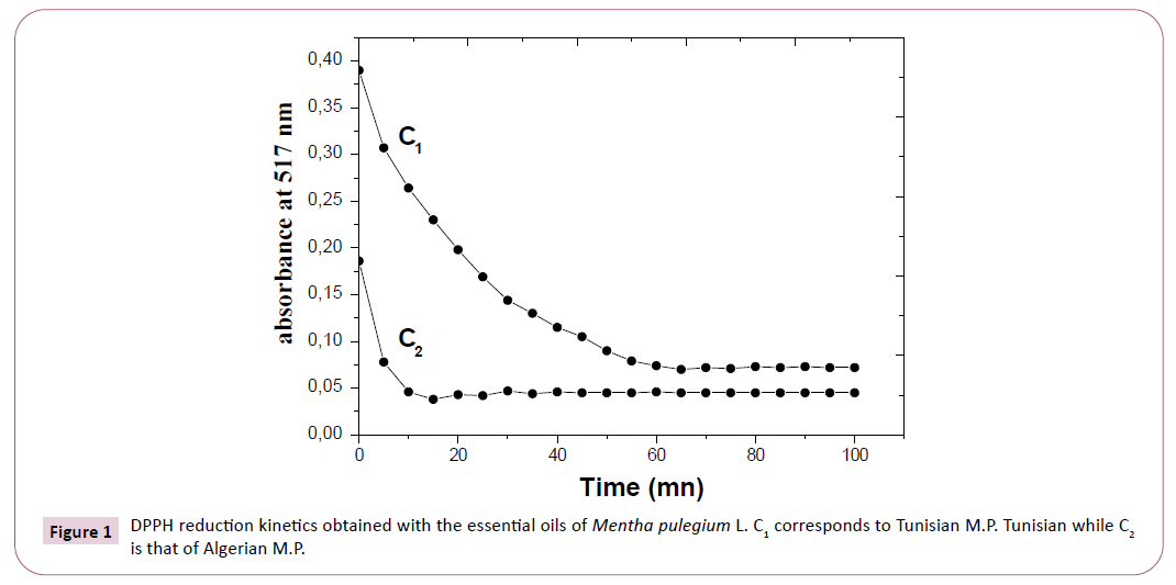
The kinetic of the antioxidant activity of essential oils is presented by the curves of Figure 1. For the essential oils examined, the reaction is biphasic, with a rapid decline in absorbance in the first minutes, followed by a slower step, until equilibrium is reached, then there are two areas:
Figure 1: DPPH reduction kinetics obtained with the essential oils of Mentha pulegium L. C1 corresponds to Tunisian M.P. Tunisian while C2 is that of Algerian M.P.
• Zone with strong kinetics of trapping of the radical observed after the first fifteen minutes for the Algerian essential oil and less fast of the order of 50 minutes for the Tunisian essential oil.
• Zone with low kinetics of trapping of the DPPH radical or zone of trend towards equilibrium for the two oils.
The results show that the reaction between DPPH and essential oils reaches equilibrium after a short time for the Algerian essential oil.
Antifungal activity: Regarding antifungal activity, we are interested in the three pathogens that are responsible for a majority of human fungal infections which are Aspergillus Niger, Candida albicans, and Cryptococcus neoformans. For each of the three pathogenic fungal species studied, MIC values essential oils have been determined.
The essential oils activity against C. neoformans is more interesting than A. Niger and C. albicans. However the results obtained show that the essential oils of the Algerian and Tunisian Mentha pulegium have significant antifungal activity.
Results of the minimum inhibitory concentrations (MIC) are summarized in Table 4.
Table 4 MIC of essential oils.
| Mentha pulegium Algeria | Mentha pulegium Tunisia | |
|---|---|---|
| A. niger MIC (ppm) | 313 | 625 |
| C. albicans MIC (ppm) | 625 | 625 |
| C. neoformans MIC (ppm) | 313 | 313 |
In Table 5 we propose some values of the minimum inhibition concentration for the three pathogens studied, we therefore present the minimum and maximum values (MIC) of certain essential oils according to (Robert L. McFeeters) [25].
Table 5 Minimum and maximum (MIC) of some essential oils according to (Robert L. McFeeters).
| A. niger MIC (ppm) | Pogostemon cablin(Indonesia): 160 | Myrtle communis (Albania): 625 |
| C. albicans MIC (ppm) | Cedrus atlantica (Morocco): 80 | Citrus junos (Japan): 1250 |
| C. neoformans MIC (ppm) | Santalum spicatum (Australia): 40 | Nymphaea caerulea (India): 625 |
The essential oils of both species have shown significant antifungal activity against the tested fungi. This bioactive power observed in the two oils is mainly attributed to their high contents in terpene phenols [38].
Antileishmanial activity: Antileishmanial properties of essential oils are described in Table 6. The IC50 values evaluated in L. major promastigote and axenic amastigote forms were around 80-100 μg.mL-1.
Table 6 In vitro antileishmanial activity results for essential oil.
| Compound | Cytotoxicity CC50 ± SD (mg.mL-1) | In vitro antileishmanial activity on L. dovani axenic amastigotes IC50 aa ± SD (mg.mL-1) | Intramacrophages amastigotes IC50 ia ± SD (mg.mL-1) |
|---|---|---|---|
| Mentha pulegium Algeria | 80 | 90 | 100 |
| Mentha pulegium Tunisia | 90 | 100 | 100 |
In the present study, the extracts of Mentha pulegium had in vitro effects on Leishmania Major and Donovani (LV9) Axenic amastigotes.
Secondary metabolites such as alkaloids, flavonoids, saponins and terpenoids were known to possess antileishmanial activities. Thus the inhibitory effect of the extracts could be due to presence of wide range of secondary metabolites with different polarities [39-41].
This activity was explained by the mechanism of extract either by killing parasites or causing metabolic disorders to inhibit the reproduction of parasites [42].
Conclusion
The present study has allowed us to describe and to compare chemical composition of Algerian and Tunisian Mentha pulegium essential oils. We have also studied antioxidant, antifungal and antileishmanial activities.
It can be concluded that the essential oils of Algerian and Tunisian Mentha pulegium seem to be a natural means for the antifungal and antileishmanial treatment and also seem good antioxidants. These essential oils can be used as an antibiotic or organic food preservative, therefore a natural replacement for harmful synthesized chemicals. However, the toxicological effects of these oils have to be investigated before human consumption before considering their use for food preservation or medicinal purposes.
References
- Kouassi EK, Ouattara S, Seguin C, Fournel S, Frisch B et al. (2018) Etudes de quelques propriétés biologiques de Ocimum Gratissimum L, laminaceae recoltee a Daloa (Cote d'Ivoire). European Scientific Journal.14: 1857-7881.
- Heath HB (1981). Source Book of Flavours. Westport: Avi 890.
- Robert G (2000) Les Sens du Parfum. Osman Eroylles Multimedia. Paris. p: 224.
- Owen RW, Giacosa A, Hull WE, Haubner R, Spiegelhalder B, et al. (2000) The antioxidant/anticancer potential of phenolic compounds isolated from olive oil. Eur Jour Cancer 36: 1235-1247.
- Hmiri S, Rahouti M, Habibi Z, Satrani B, Ghanmi M, et al. (2011) Evaluation du potential antifongique de Mentha pulegium et d'eucalyptus camaldulensis dans la lute biologique contre les champignons responsables de la deterioration des pommes en conservation. Bulletin de la Société Royale des Sciences de Liège 80: 824-836.
- Moleyar V, Narasimham P (1986) Antifungal activity of some essential oil components. Food Microbiology 3: 331-336.
- Soliman KM, Badeaa RI (2002) Effect of oil extracted from some medicinal plants on different mycotoxigenic fungi. Food Chem Toxicol 40: 1669-1675.
- Magina MDA, Dalmarco EM, Wisniewski A, Simionatto EL, Dalmarco JB, et al. (2009) Chemical composition and antibacterial activity of essential oils of Eugenia species. J Nat Med 63: 345-350.
- Bouzouita N, Kachouri F, Ben Halima M, Chaabouni MM (2008) Composition chimique et activités antioxydant, antimicrobienne et insecticide de l’huile essentielle de Juniperus Phoenicea. J Soc Chim Tunis 10: 119-125.
- Bruneton J (1993) Pharmacognosie Phytochimie Plantes médicinales. 2nd edition. Lavoisier, Paris. 41-54.
- Cremieux A (1990) Etude de six huiles essentielles: composition chimique et activité antibactérienne. Study of six essential oils: chemical composition and antibacterial activity. J Food Science 85: 2437-2444.
- Bhaskara Reddy MV, Angers P, Gosselin A, Arul J (1997) Characterization and use of essential oil from Thymus vulgaris against Botrytis cinerea and Rhizopus stolonifer in strawberry fruits. Phytochemistry 47: 1515-1520.
- Tzortzakis NG (2006) Maintaining postharvest quality of fresh produce with volatile compounds. Innovative Food Sci Emerg Technol 8: 111-116.
- Amarti F, Satrani B, Ghanmi M, Farah A, Aafi A, et al. (2010) Composition chimique et activité antimicrobienne des huiles essentielles de Thymus algeriensis Boiss. and Reut et Thymus ciliatus (Desf) Benth. du Maroc, Biotechnol. Agron Soc Environ 14: 141-148.
- Mahboubi M, Haghi G (2008) Antimicrobial activity and chemical composition of Mentha pulegium L. essential oil. J Ethnopharmacol 119: 325-327.
- Maihebiau P (1994) La nouvelle aromathérapie: biochimie aromatique et influence psychosensorielle des odeurs. Lausanne 635.
- Bouyahya, Et-Touys A, Bakri Y, Fellah H, Abrini J, et al. (2017) Chemical composition of Mentha pulegium and Rosmarinus officinalis essential oils and their antileishmanial, antibacterial and antioxidant activities. PII: S0882-4010(17): 30581-8.
- Ahmed H, El-Ghorab (2006) The Chemical Composition of the Mentha pulegium L. Essential Oil from Egypt and its Antioxidant Activity. Journal of Essential Oil Bearing Plants 9 (2): 183-195.
- Abdellia M, Moghrani H, Aboun A, Maachi R (2016) Algerian Mentha pulegium L. leaves essential oil: Chemical composition, antimicrobial, insecticidal and antioxidant activities. Industrial Crops and Products 94: 197-205.
- Sheille R, Mondello L, Marriott P, Dugo G (2002) Characterization of lavender essential oils by using gas chromatography-mass spectrometry with correlation of linear retention indices and comparison with comprehensive two-dimensional gas chromatography. Journal of Chromatography A 970: 225-234.
- Adams RP (2007) Identification of Essential Oil Components by Gas Chromatography/Mass Spectrometry.
- Belboukhari N, Merzoug Z, Cheriti A, Sekkoum K, Yakoubi M (2013) Etude de la caractérisation et la composition qualitative des huiles essentielles de six plantes médicinales par spectroscopie IR. Phyto Chem & BioSub Journal 7(1).
- Braca A., Sortino C, Politi M, Morelli I, Mendez J (2002) Antioxidant activity of flavonoids from Licania licaniaeflora. J Ethnopharmacol 79: 379-381.
- Tyler H, Jones, Erin E, McClelland, McFeeters H, et al. (2017) Novel Antifungal Activity for the Lectin Scytovirin: Inhibition of Cryptococcus neoformans and Cryptococcus gattii. Frontiers in Microbiology 8: 755.
- Chelsea N, Powers, Prabodh Satyal, John A, Mayo, et al. (2019) Bigger Data Approach to Analysis of Essential Oils and Their Antifungal Activity against Aspergillus Niger, Candida albicans, and Cryptococcus neoformans. Molecules 24: 2868.
- Chelsea N, Powers Jessica L, Osier Robert L, McFeeters, Brianne Brazell C, et al. (2018) Antifungal and Cytotoxic Activities of Sixty Commercially-Available Essential Oils. Molecules 23: 1549.
- Philippe M, Loiseau, Gupta S, Verma A, Srivastava S, et al. (2011) In Vitro Activities of New 2-Substituted Quinolines against Leishmania donovani. Antimicrobial Agents and Chemotherapy 55: 1777-1780.
- Campos Vieira N, Vacus J, Fournet A, Baudouin R, Bories C, et al. (2011) Antileishmanial activity of a formulation of 2-n-propylquinoline by oral route in mice model. Activite antileishmanienne d’une formulation de 2-n- propylquinoleine administree par voie orale chez la souris. Parasite 18: 333-336.
- Uwineza MS, El Yousfi B, Lamiri (2018) Activités antifongiques in vitro des huiles essentielles de Mentha pulegium, Eugenia aromatica et Cedrus atlantica sur Fusarium culmorum et Bipolaris sorokiniana. Antifungal activities of essential oils of Mentha pulegium, Eugenia aromatica and Cedrus atlantica on Fusarium culmorum and Bipolaris sorokiniana in vitro. Revue Marocaine de Protection des Plantes 12: 19-32.
- Amaral Mônaco Foganholi AP, Feijó Souza Daniel J, Santiago DC, Orives JR, Pereira JP, et al. (2015) Chemical composition and antifungal activity of pennyroyal essential oil in different stages of development. Composicao qulmica e atividade antifúngica do oleo essential de poejo em differences estagios de desenvolvimento. Ciencias Agrarias, Londrina. 36: 3091-3100.
- Kamkar A, Javan AJ, Asadi F, Kamalinejad M (2010) the ant oxidative effect of Iranian Mentha pulegium extracts and essential oil in sunflower oil. Food and Chemical Toxicology 48: 1796-1800.
- Stoyanova A, Georgie V, Kula J, Majda T (2005) Chemical composition of the essential oil of Mentha pulegium from Bulgaria. Journal of Essential Oil Research 17: 475-477.
- Lorenzo D, Paz D, Dellacassa E, Davies P, Vila R, et al. (2002) Essential oils of Mentha pulegium and M. rotudifolia from Uruguay. Brazilian Archives of Biology and Technology 45: 519-524.
- Teixeira B, Marques A, Ramos C, Batista I, Serrano C, et al. (2012) European pennyroyal (Mentha pulegium) from Portugal : Chemical composition of essential oil and antioxidant and antimicrobial properties of extracts and essential oil. Industrial Crops and Products 36: 81-87.
- Yasa H, Çelik Onar H, Yusufoglu AS (2012) Chemical Composition of the Essential Oil of Mentha pulegium L. from Bodrum, Turkey. Journal of Essential Oil Bearing Plants 15: 1040- 1043.
- Asma E, Hervey C, Emmanuelle V, Belgacem H, Karim H (2018) Assessing the fatty acid, essential oil composition, their radical scavenging and antibacterial activities of Schinus terebinthifolius Raddi leaves and twigs. J Food Sci Technol 55: 1582-1590.
- Brahmia F, Abdenourb A, Brunoc M, Silviac P, Alessandrac P, et al. (2016) Chemical composition and in vitro antimicrobial, insecticidal and antioxidant activities of the essential oils of Mentha pulegium L. and Mentha rotundifolia (L.) Huds growing in Algeria. 88: 96-105.
- Eajjouri M (2008) Activité antifongique des huiles essentielles de Thymus bleicherianus Pomel et Thymus capitatus (L.) Hoffm, Link contre les champignons de pourriture du bois d’oeuvre. Biotechnol Agron Soc Environ 12: 345-351.
- Chan-Bacab MJ, Pena-Rodriguez LM (2001) Plant natural products with leishmanicidal activity. Nat Prod Rep 18: 674-688.
- Salehabadi A, Karamian M, Farzad MH, Namaei MH (2014) Effect of root bark extract of Berberis vulgaris L. on Leishmania major on Balb/c mice. Parasitol Res 113: 953-957.
- Ogungbe IV, Setzer WN (2013) In-silico Leishmania target selectivity of ant parasitic terpenoids. Molecules 18: 7761-7847.
- Ahmet O, Cenk D, Husniye K, Hatice E, Cumhur G, et al. (2014) Antileishmanial Activity of Selected Turkish Medicinal Plants. Tropical Journal of Pharmaceutical Research 13: 2047-2055.
Open Access Journals
- Aquaculture & Veterinary Science
- Chemistry & Chemical Sciences
- Clinical Sciences
- Engineering
- General Science
- Genetics & Molecular Biology
- Health Care & Nursing
- Immunology & Microbiology
- Materials Science
- Mathematics & Physics
- Medical Sciences
- Neurology & Psychiatry
- Oncology & Cancer Science
- Pharmaceutical Sciences

