Comparative Anatomy of the Lower Respiratory Tract of Domestic Fowl (Gallus gallus domesticus) and Guinea Fowl ( Numida meleagris): A Histological Study
1Department of Veterinary Anatomy, Usmanu Danfodiyo University, Sokoto, Nigeria
2Department of Theriogenology and Animal production, Usmanu Danfodiyo University, Sokoto, Nigeria
3Department of Veterinary Pathology, Usmanu Danfodiyo University, Sokoto, Nigeria
- *Corresponding Author:
- Bello A
Department of Veterinary Anatomy
Usmanu Danfodiyo University
Sokoto, Nigeria
Tel: 8039687589
E-mail: abccrcfge28@gmail.com
Received date: March 12, 2018; Accepted date: March 19, 2018; Published date: March 26, 2018
Citation: Bello A, Shehu SA, Sununu AT, Umaru MA, Umar AA, et al. (2018) Comparative Anatomy of the Lower Respiratory Tract of Domestic Fowl (Gallus gallus domesticus) and Guinea Fowl (Numida meleagris): A Histological Study. J Anat Sci Res. Vol.1 No.1:3
Copyright: © 2018 Bello A, et al. This is an open-access article distributed under the terms of the Creative Commons Attribution License, which permits unrestricted use, distribution, and reproduction in any medium, provided the original author and source are credited.
Abstract
A study aiming at investigating the differences in the histological structures of the lower respiratory tract of guinea fowl and domestic fowl was conducted in the department of Veterinary Anatomy of Usmanu Danfodiyo University, Sokoto. A total of twenty four adult birds (12 domestic fowls and 12 guinea fowls) without sex consideration were sacrificed, after which, the lower respiratory tract (trachea and lungs) were identified based on their positions, shapes and relations with other thoracic organs. The relations of the trachea with the neck in the samples were recorded. The lower respiratory tract of each bird was dissected out and fixed in 10% formalin. The samples were subjected to the routine histological processes which ultimately led to the production of histological slides of the various organs. The sides showed that the trachea of both species was lined by respiratory epithelium (pseudo stratified ciliated columnar epithelium) with simple branched tubular mucous glands and goblet cells. Lamina propria-submucosa of the trachea was supported by hyaline cartilage in all the species, which show clear morphological variation by been thicker (double layer) and comprise of loose connective tissue, with large bundles of collagen fibers in guinea fowl than in domestic fowl with single thin layer and comprise of loose connective tissue, with little bundles of collagen fibers. The lungs of the two species appeared to be the same with significant difference in the presence of connective tissue fibers in the lungs of the guinea fowl when compared to the lungs of the domestic fowl. Based on this finding it shown that there is significant difference between the lower respiratory tract of guinea fowl and domestic fowl histologically.
Keywords
Comparative anatomy; Lower respiratory tract; Gallus gallus domesticus; Numida meleagris domesticus
Introduction
Poultry refers to all domesticated birds kept for egg or meat production [1-3] and these include chicken, turkey, ducks, geese, pigeon and guinea fowl [1,4-7]. Also the birds reared or hunted for a purpose are members of bird group collectively known as poultry [4,8]. Functionally, birds also serve as experimental animals [9], household pets [10,11] and as a major source of animal protein [12,13]. In the past, poultry keeping was a sideline occupation, gradually it developed into a commercial enterprise involving millions of birds. Globally, poultry has become the cheapest and richest source of animal protein among the various livestock, hence one of the most important branches of livestock industry [7,14,15].
Poultry management systems in Africa are differentiated on the basis of flock size and input-output relationships [16]. These include extensive, semi-intensive and intensive management systems [17]. In the extensive management system, different species of poultry that include guinea fowls, chickens, ducks and turkeys are kept. Chickens are type of domesticated fowl, a subspecies of the red jungle fowl [17] and they are one of the most common and wide spread domestic animals. Chickens are birds kept for both meat and egg, and are raised for meat as broiler chickens, whereas chickens raised for egg are called layers [8,18]. Poultry meat is appreciated and acclaimed as being the favorite worldwide. The meat is wholesome, has low fat, easy to prepare, tender and loved by most people [7]. There is a belief that chicken is good for children and it assist in brain development and subsequently intelligence [14]. There are approximately 8,900 species of living birds in the world [19]. Chickens will naturally live for 6 or more years, but broiler chickens typically take less than 8 weeks to reach slaughter size, while a free range or organic meat chicken will usually be slaughtered at about 14 weeks of age [20].
Productively chicken are of two types namely; domestic and feral chickens [21]. Feral chickens (domesticated birds that have reverted to a wild state) have been observed to eat a wide variety of plant matter, including berries, seeds, and grasses, availing themselves of all food sources in their habitats [3]. They scratch the forest floor for insects and snails, and they snatch figs and other fruits from the trees [2,3,20]. Chickens have even been observed forming symbiotic relationships with cattle, by pecking at the flies that swarm around the cows’ faces and by scavenging on their waste [20]. They have also been observed to eat carrion [3]. The basic behavioral repertoire of domestic chickens is fundamentally the same as that of their wild ancestors [19].
Guinea fowls (Numida meleagris) originated in Africa and were first domesticated by ancient Egyptians [1,3,9]. They are currently being reared in many parts of the world. In countries such as Egypt and India, guinea fowls are now produced commercially on a large scale, while in some African countries which include Nigeria, Malawi and Zimbabwe, guinea fowl production is in its in f ancy [3,8,14]. Ho w e v er , N. meleagris (red wattled guinea fowl) is an origin of West Africa and observed to be a docile bird that can lay in captivity [8].
The respiratory system generally consists of upper respiratory tract (nostril, nasal cavity and pharynx) and lower respiratory tract (larynx, trachea, syrinx and lungs) that allow for a union between air and blood with the aim of exchange of gases [21,22]. In addition, the major functions of the respiratory system are oxygen and carbon dioxide exchange, and balance in body temperature [23]. Air inspired during respiration passes from the nasal cavity to the larynx and continues via the trachea and enters the syrinx and bronchi [24].
There have been a lot of studies related to anatomy and physiology of the lower respiratory tract of various domestic [3,6,15,21,22,25-27] and wild bird in the world [1,2,5,10,13,14,20], there is death of information on the comparative lower respiratory tract of domestic fowl and guinea fowl in this part of the country (Sokoto, Nigeria). Although the anatomical structures of domestic animals are basically the same except for minor differences in shape, size and structure, there is still the need to study the comparative anatomical parts different animal species of different generic group to note their normal similarities and differences in shape, size, location and composition, grossly and histologically. The aim of this work is to study the comparative histological difference of the lower respiratory tract of domestic fowl and guinea fowl in Sokoto Nigeria.
Materials and Methods
The study was conducted in Sokoto metropolis, the capital of Sokoto state of Nigeria. Sokoto state geographically was located on 12º15N and 05ºE latitude, and 38 m above sea level. The state has savannah vegetation of short grasses in the south and thorn in the north. Sokoto was bounded by Zamfara state to the east, Niger Republic to the north and Kebbi state to the west and southwest [28].
The state was ranked second in the nation livestock population with an estimated of 34,532 horses, 51,388 donkeys. Two million cattles, one million sheeps, two million goats, four million chickens [8].
Animal sample and source
A total of twenty-four adult birds (12 domestic fowls and 12 guinea fowls) were used for this study. The birds were purchased from the Sokoto metropolitan central market of Sokoto State. They included 12 domestic fowls (6 cocks and 6 hens) and 12 guinea fowl (6 cocks and 6 hens). They were transported to the Histology unit (Anatomy Laboratory) of the Department of Veterinary Anatomy, Usmanu Danfodiyo University, Sokoto.
Sample processing and dissection
The animals were acclimatized and observed before euthanizing for one week (7 days) to ascertain the health status of the birds. The life birds were euthanized with chloroform, dipped in tap water and placed on the dissecting table. Defeathering of the entire carcass was done to give a better accessibility of visceral organs for clean dissection. A sharp dissecting scissors was used to cut through the abdominal wall along the posterior margin of the sternum and carefully reflected by cutting further through the thoracic region exposing the thoracic and abdominal visceral organs. The respiratory system was then studied in situ and its relation with other structures was noted. Then the visceral organs were dissected out, this was done by cutting gastrointestinal tract at the termination of the esophagus, the proventriculus, gizzard, the pancreas which rest on duodenal loop, and the large intestine including cecae are removed after detailed examination. With the aid of scissors a cut is made through the uppermost commissure of the beak. Further, cut through the larynx to open the trachea throughout its length, followed by the bronchi into the lungs were made [12].
Gross observation
The lower respiratory tract (comprising of the trachea, bronchus and lungs) samples were observed grossly for relation with other structures, shape, size, and number of rings of trachea of the two species.
Histological techniques
Tissue samples were taken from the cervical, medical and thoracic regions of the trachea and were fixed in 10% neutral buffered formalin for 7 days. The tissue was then processed as per method adapted by Bello et al. [25]. The paraffin blocks formed were sectioned into 5 μm thickness using manual serial microtome, and stained using haematoxylin and eosin (H&E) for normal morphological viewing with microscope [12,25]. The sections were observed using a light microscope and micrographs were taken using a common digital camera with 12.1 mega pixels.
Results and Discussion
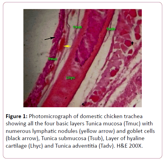
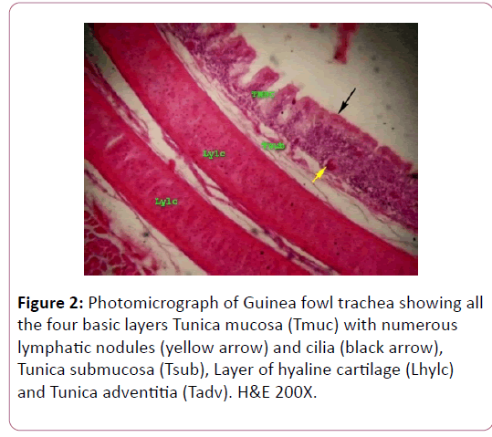
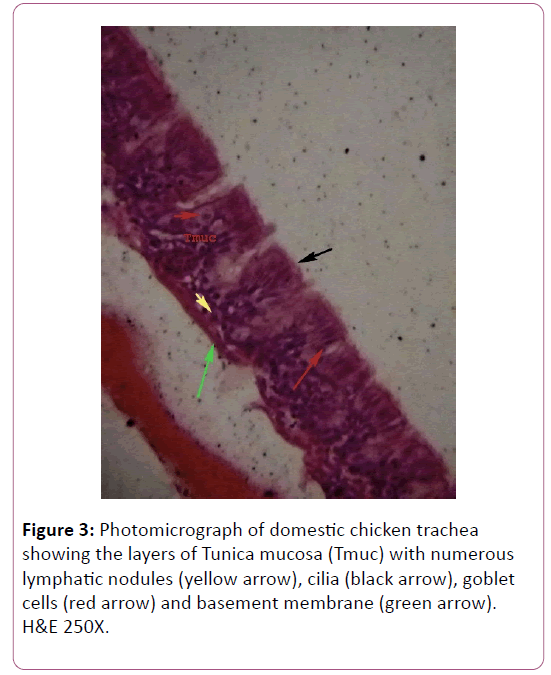
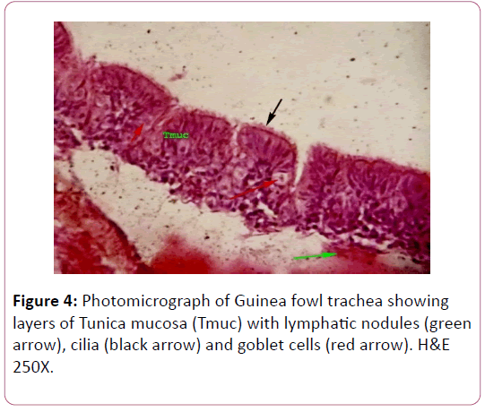
The study shows that the trachea of domestic fowl and guinea fowl consists of mucosa, sub mucosa and adventitia (Figures 1 and 2). The mucosal layer of domestic and guinea fowl are lined with pseudo stratified ciliated columnar epithelium with oval nuclei with presence of intraepithelial goblet cells. There are high numbers of mucous glands in the guinea fowl than in the domestic fowl trachea and becomes fewer or even absent in the posterior/ caudal region of the domestic fowl trachea (Figures 3 and 4). In guinea fowl the mucosal layer has numerous crypts lined with pseudo stratified ciliated columnar epithelium which is housed with numerous goblet cells where its nuclei near the surface, the glands are simple alveolar mucous glands (Figure 4). This findings (the trachea of domestic fowl is lined by stratified columnar ciliated cells rich with goblet cells) agreed with in their review on Guinea fowl, Duck and Goose respectively [5,10,29,30]. Al-Mussawy, in their study on Japanese quail, on study of Turkey and Ayeni and in the study of goose also confirms similar observations [2,21]. This may be because ciliary movement by a mucus layer (mucociliary system) is believed to perform a cleaning function by removing foreign bodies (bacteria, virus, dust etc.) from the respiratory tract [21,30,31].
Figure 1: Photomicrograph of domestic chicken trachea showing all the four basic layers Tunica mucosa (Tmuc) with numerous lymphatic nodules (yellow arrow) and goblet cells (black arrow), Tunica submucosa (Tsub), Layer of hyaline cartilage (Lhyc) and Tunica adventitia (Tadv). H&E 200X.
Figure 3: Photomicrograph of domestic chicken trachea showing the layers of Tunica mucosa (Tmuc) with numerous lymphatic nodules (yellow arrow), cilia (black arrow), goblet cells (red arrow) and basement membrane (green arrow). H&E 250X.
Figure 4: Photomicrograph of Guinea fowl trachea showing layers of Tunica mucosa (Tmuc) with lymphatic nodules (green arrow), cilia (black arrow) and goblet cells (red arrow). H&E 250X.
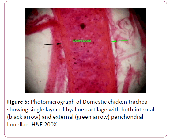
In domestic fowl and guinea fowl, the epithelium was observed to lies on basement membrane contains basal cells which have flattened nuclei that separate it from the underlying lamina propria as shown in (Figures 2 and 3). Result shown that, in domestic fowl lamina propria is thin consists of loose connective tissue, and heavy accumulation of connective tissue fibres, while in guinea fowl lamina propria consists of loose connective tissue and fibers, with presence of blood vessels, and sub mucosa that consists of complete cartilaginous rings as shown in (Figures 5 and 6).
This is in agreement with the observations of Maina and Nathanel and Onuk et al. who observed obvious loose connective tissue and heavy accumulation of connective tissue fibres in the lamina propria mucosa of the ostrich and goose respectively [11,31]. In guinea fowl mucosal layer has very clear crypts lined with respiratory epithelium which houses numerous of goblet cells is in agreement with Yildizi et al. findings that intraepithelial mucous glands are abundant in the trachea of flightless birds. Yildizi et al. also shown that the Lamina propria of coot birds has moderate loose connective tissue with heavy accumulation of lymphocyte, this observation is in agreement with our finding in guinea and show large bundles of collagen fibers and numerous aggregations of lymphocytes in domestic fowl [15].
The submucosal layer is made of cartilage which is thicker in guinea fowl than that of domestic fowl. The later were single and contains scattered of lacunae which consists of chondrocyte as shown in (Figure 5). In guinea fowl, the cartilage is also complete ring but there are doubled cartilagenous layer with prominent interchondral membrane as shown in (Figure 2). Cartilage thickness is thicker in guinea fowl than of domestic fowl. The above findings were contrary to those reported by Kaites et al. which showed the presence of single cartilagenous layer with no visible demarcation in wild guinea fowl [26].
The last layer is adventitia which contains loose connective tissue, blood vessels and nerves in guinea and domestic fowl consists of clear nerve fascicles with blood vessels as shown in (Figures 3 and 4), is in agreement with Yildizi et al. [15], Maina and Nathanel [11], Onuk et al. [29] and Kaites et al. [26].
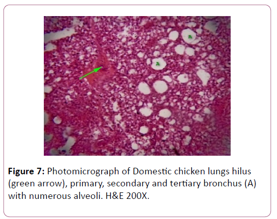
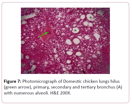
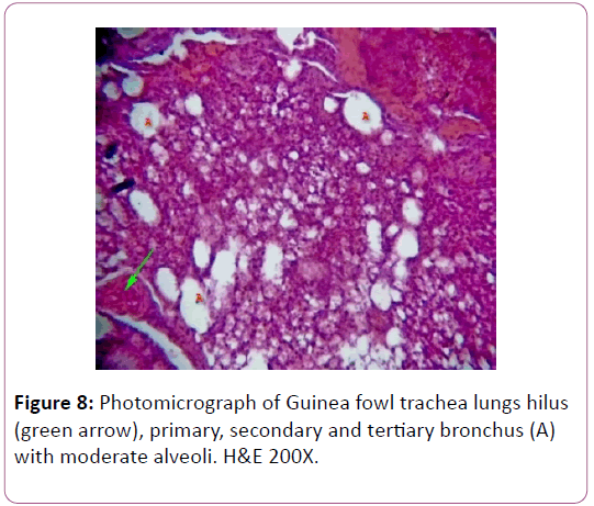
Result have shown that, in both guinea and domestic fowl, the lungs appeared to be histological identical except that, the presence of much connective tissue in the lungs of the guinea fowl which show more intense than in the lungs of the domestic fowl (Figures 7 and 8).
Conclusion and Recommendations
Based on the above findings, it showed that there is histological similarities between domestic fowl and guinea fowl lower respiratory tract (trachea and lung) histologically with much difference in the cartilagenous ring between the two species. The information obtained in this study will serve as a base-line data in compiaring the two species anatomical and physiologically in area of research and teaching purrpose.
References
- Anon (1976) How to improve the performance of local chicken, Extension Guide, ABU Zaria 79: 1-7.
- Anonymous (1998) Domesticating and raising of guinea fowl on free range. Small livestock and wildlife. Farming World 24: 25-26.
- Ayeni, JSO, (1980) The biology and utilization of the Helmet Guinea Fowl (Numida galeata pallas) in Nigeria. Ph.D. Thesis, University of Ibadan.
- Baumel JJ, King SA, Breasile JE, Evans HE, Berge JCV (1993) Handbook of Avian Anatomy (Nomina Anatomica Avium). Publications of the Nuttall Ornithological Club, Cambridge. Massachusetts pp: 257-299.
- Binali W, Kanengoni E (1998) Guinea fowl production. A training manual board on science and technology for international development. Washington, National Academy Press pp: 115-125.
- Dyce KM, Sack WO, Wensing CJ (2010) Textbook of Veterinary Anatomy (4th Edn) Sounders Publishers pp: 794-812
- Tye GA, Gyawu P (2001) The benefits of intensive guinea fowl production in Ghana. World Poul 17: 53-54.
- Knox I (2000) Guinea fowl. Farm Diversification Information Service.
- Betty J (1979) Domesticated chicks and geese pp: 112-82.
- Demirkan AC, Haziroglu RM, Kurtul I (2006) Air sacs (Sacci pneumatici) in mallard ducks (Anas platyrhynchos), Ankara Univ. Vet Fak Derg 53: 75-78.
- Maina JN, Nathanel C (2001) A qualitative and quantitative study of the lung of an Ostrich (Struthio camelus). J Exp Biol 204: 2313-2330.
- Ojo SA (1990) Guide to the study and dissection domestic fowl. ABU Press, Zaria.
- Schmidt E, Robert (2013) The avian respiratory system, USA. Western veterinary conference pp: 1-15.
- Oluyemi JF, Robert FA (2007) Poultry production in the warm wet climates (2nd Edn) Ibadan. Golden Wallet Press pp: 2.
- Yildizi H, Yilmaz B, Arican I (2005) Morphological structure of the syrinx in the Bursa roller pigeon (Columba livia), Bull Vet Inst. Pulawy 49: 323-327.
- King AS, Mclelland J (1975) Outlines of avian anatomy, London, Bailiere Tindall pp: 1-42.
- Mohammad RFE (2004) Fundamentals of the histology of birds part I: Histology of Poultry: An Introductory Text for Veterinary Students (2nd Edn) Research gate publishers pp: 36-54.
- Morony JJ, Bock WJ, Farrand J (1975) Reference list of the birds of the world. American museum of natural history, Department of Ornithology, New York.
- Appleby MC, Mench JA, Hughes BO (2004) Poultry Behaviour and Welfare. Wallingford, U.K., CABI Publishing.
- Belshaw R. H, (1985). Guinea fowl of the world “world of ornithology”. Minirod Book Services, Hampshire, England.
- Mussawy AL, (2011) Anatomical and histological study of major respiratory organs (Larynx, Trachea, Syrinx, Bronchi and Lungs) in indigenous male Turkey (Meleagris gallopava). M.Sc. Thesis, AL-Qadisiya Uniersity, Vet. Med. College.
- Kabak M, Orhan IO, Haziroglu RM (2007) The gross anatomy of larynx, trachea and syrinx in the long-legged buzzard (Buteo rufinus). Anat Histol Embryol 36: 27-32.
- King AS, Baumel JJ, King AS, Lucas AM, Breazile JE, et al. (1979) Systema respiratorium in Nomina Anatomica Avium, London. Academic Press pp: 227-265.
- Corral JPD, LM Pastor (1995) Anatomy and histology of the lung and air sacs of birds in histology, ultrastructure and immunohistochemistry of the respiratory organs in non-mammalian vertebrtaes. Murcia, Spain, Publications of the University of Murcia pp: 179-233.
- Bello A, Onyeanusi BI, Sonfada ML, Adeyanju JB, Umaru MA, et al. (2012) Histomorphological studies of the prestiatal development of oesophagus of one hump camel (Camelus dromedaries). Scient J Agricul 1: 100-104.
- Kaites T, Alisteair F (1986) The Complete book of raising livestock and poultry: A smallholder's guide, London, Martin Dunitz pp: 9-10
- Reece WO (2005) Avian respiratory system morphology. In: Function Anatomy and Physiology of Domestic Animals (3rd Edn) Lippincott Williams and Wilking pp: 230-268.
- Sokoto BA (2001) The Study of local geography of Nigerian secondary school (1st Edn) Sokoto, Ministry of Education pp: 104-110
- Onuk B, Haziroglu RM, Kabak M (2010) The gross anatomy of larynx, trachae and syrinx in Goose (Anser anser domesticus), Kafkas Univ. Vet Fak Derg 16: 443-450.
- Al-Mamoori NAM, Al-Ghakany SSA (2015) Anatomical and morphometric study of the trachea in Bee-eater Bird (Merops orientalis). J Agricul Vet Sci 8: 58-61.
- Aughey E, Frye FL (2001) Comparative Veterinary Histology with Clinical Correlates, London. Manson Ltd pp: 93-94.
Open Access Journals
- Aquaculture & Veterinary Science
- Chemistry & Chemical Sciences
- Clinical Sciences
- Engineering
- General Science
- Genetics & Molecular Biology
- Health Care & Nursing
- Immunology & Microbiology
- Materials Science
- Mathematics & Physics
- Medical Sciences
- Neurology & Psychiatry
- Oncology & Cancer Science
- Pharmaceutical Sciences
