ISSN : 2576-392X
Dentistry and Craniofacial Research
Case Report of Missing Isthmus Endodontic Mishap
Badria Al-Matrafi1*, Reem Al-Idrisi1, Hussein Mukhles2, Hisham Zaidan2, Abdulmajeed Al-Takhees2, Al-Bishi Dalal3, Saud Al-Saif4, Amal alshehr5, Norah Alotaibi6 and Mshael Almohaimel6
1Department of Dentistry, BDS, KSU, AGD, University of South California, United State of America, ARD, SCFHS, Consultant restorative Prince Sultan Military Medical City, Saudi Arabia
2Department of Dentistry, Consultant Endontist PSMMC, Riyadh, Saudi Arabia
3Department of Dentistry, GP Ministry of Health, Riyadh, Saudi Arabia
4Department of Dentistry, Riyadh Elm University, Riyadh, Saudi Arabia
5Department of Dentistry, Dental Hygienist PSMMC, Riyadh, Saudi Arabia
6Department of Dentistry, Princess Nourah bint Abdulrahman University, Riyadh, Saudi Arabia
- *Corresponding Author:
- Badria Almatrafi
Department of Dentistry,BDS, KSU, AGD,
University of South California,
United State of America,ARD,SCFHS,
Consultant restorative Prince Sultan Military Medical City,
Saudi Arabia,
E-mail: dr_badria@hotmail.com
Received date: October 13, 2023, Manuscript No. IPJDCR-23-18128; Editor assigned date: October 16, 2023, PreQC No. IPJDCR-23-18128 (PQ); Reviewed date: October 30, 2023, QC No. IPJDCR-23-18128; Revised date: November 06, 2023, Manuscript No. IPJDCR-23-18128 (R); Published date: November 13, 2023, DOI: 10.36648/2576-392X.8.3.147
Citation: Al-Matrafi B, Al-Idrisi R, Mukhles H, Zaidan H, Al-Takhees A, et al. (2023) Case Report of Missing Isthmus Endodontic Mishap. J Dent Craniofac Res Vol:8 No:3
Abstract
The primary objective of endodontic treatment is the comprehensive removal of diseased pulp tissue from the root canal system. This involves shaping and cleaning the canal, followed by filling it with inert material, effectively reducing or eliminating the risk of re-infection and thereby ensuring treatment success. Deviation from accepted clinical standards can result in treatment failure, particularly in cases of persistent apical periodontitis following conventional root end surgery.
The anatomical complexity within the root canal system includes a thin passage called an isthmus, housing pulp or tissue originating from the pulp. It is essential to recognize that the isthmus is not an independent entity but an integral component of the root canal system. The isthmus must be thoroughly cleaned, shaped, and filled. Endodontists need to be cognizant of the presence of isthmuses, especially in molars and premolars, located at the 3 millimeter level from the apex, occurring in approximately 80%-90% of instances requiring apical surgery.
The intricate nature of isthmus preparation contributes to a significantly lower success rate in teeth with isthmuses compared to those without. Teeth with existing isthmuses require greater weakening to create space for both canals, posing a challenge to the overall success of the endodontic procedure. Addressing this complexity is crucial for improving treatment outcomes and ensuring the long-term success of endodontic interventions
Keywords
Isthmus; Missing shape; Endo treatment; Root end surgery; Disease pulp tissue; Failure endo treatment; Thin ribbon shape; Accepted clinical standers; Inert material root; Canal system; Accessory canal; Micro surgery; Passageway between two canal; Success rate
Introduction
Endodontic therapy aims to completely remove all diseased pulp tissue from the root canal system, clean it, and shape it so that it may be filled with an inert substance, reducing or eliminating the possibility of reinfection. Failure occurs though when endodontic therapy strays from accepted clinical standards [1].
For the treatment of persistent apical periodontitis after nonsurgical endodontic treated teeth the endodontic root-end surgery is indicated. And to make the procedure predictable, safer, and easier to perform various surgical techniques were introduced [2].
Over the past several years, there have been significant technological and procedural advancements in the area of endodontics. The manner surgery is carried out may be the one aspect of endodontics that has seen the most advancement. The distance between biological ideas and the capacity to produce consistently positive therapeutic results has shrunk because to the employment of cutting-edge equipment, novel and enhanced materials, and a surgical operating microscope. Endodontic microsurgery is the term now used to describe the use of these methods [3].
A thin, ribbon-shaped passageway between two root canals called an isthmus contains pulp or tissue deriving from pulp. The isthmus is not an independent entity but a component of the canal system. As a result, it has to be cleaned, shaped, and filled completely. The endodontist should be aware that isthmuses are present in premolars and molars at the 3 mm level from the apex in about 80% to 90% of instances when undertaking apical surgery [4]. And the incidence of isthmus failure in mandibular first molar as shown in von Arx T, et al. study was 83%.
Case Presentation
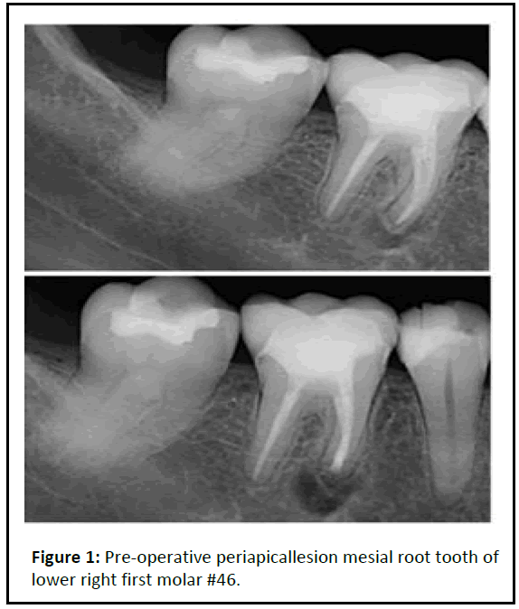
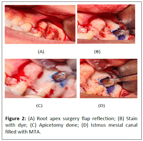
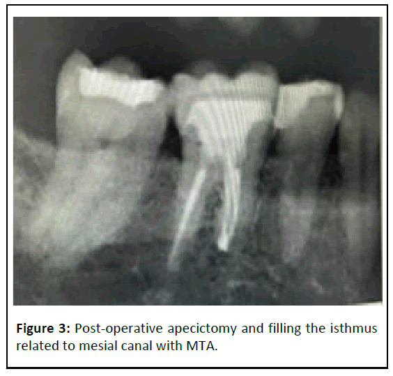
38 years old female, medically fit, came to Princes Sultan Military Medical City (PSMMC) primary care dental center complaining from slightly discomfort related to lower right area when blending her foundation makeup by beauty blender. After examination tooth #46 is endodontically treated tooth 10 years ago with excellent obturation and there is Symptomatic Apical Periodontist (SAP). The endodontist decided to do exploratory surgery to check the reason. He found missing apical isthmus related to mesial canal. Apicectomy done and the isthmus closed with MTA (Figures 1-3).
Discussion
The intricate anatomical nature of the root canal is one of the root canal treatment's challenges. Anatomic complexity is made up of many different components, such as the fin and lateral canal, and isthmuses in particular present difficulties for endodontists [5]. No precise technique for cleaning and shaping an isthmus has yet been developed, despite the fact that numerous preparation and irrigation procedures have been presented to get around the anatomical complications. The dental operating microscope, ultrasonic, modern microsurgical equipment, and biocompatible root-end filling materials are some of the technological advancements used in modern microsurgical periradicular surgery, which has produced highly successful treatment outcomes.
The increased success rates were attributed to a superior surgical site examination and the exact preparation of root-ends with micro-instruments using high magnification and improved illumination. In order to create a cavity that can be effectively filled during root end resection surgery, root-end preparation tries to remove filling material, irritants, necrotic tissue, and residues from the canals and the isthmus. The optimal rootend preparation is a class I cavity with walls parallel to and inside the anatomic shape of the root canal space that extends at least 3 mm into the root dentin. The prevalent practice in conventional surgical procedures, the use of rotating burs in a micro-handpiece, is no longer able to provide this therapeutic need. The brand or kind of tip is not clinically significant for an effective ultrasonic preparation, but rather how the tip is used. The secret to successful ultrasonic preparation is to repeatedly apply very little pressure. A softer touch improves cutting efficiency, whereas continuous pressure, as that applied by a handpiece, reduces cutting effectiveness. This is so that ultrasound can operate through vibration rather than pressure.
To increase the success rates of endodontic microsurgery for posterior teeth, an isthmus must be identified and treated. A missed isthmus, can result in failure [6]. According to Sunil Kim, et al., isthmus-present teeth had a worse success rate for endodontic microsurgery than isthmus absent teeth did. The complexity of the isthmus preparation method itself is one of the causes of the significantly lower success rate in teeth with isthmus presence. In comparison to teeth with an absent isthmus, teeth with an extant isthmus must be more weakened in order to create space for a root canal. This lowers the success rate of the surgery [7]. At the short-term follow-up after one year and the long-term follow-up after five to seven years, the clinical success of cases treated with microsurgery is reported to be as high as 96.8% and 91.5%, respectively. Similar findings have been revealed by recent prospective studies with longterm follow-up [8].
Conclusion
Even with good endodontic treatment missing isthmus can be found and cause failure of treatment. As studies shown microsurgery is predictable and effective treatment for this mishap.
References
- Siqueira JF Jr (2001) Aetiology of root canal treatment failure: Why well‐treated teeth can fail. Int Endod J 34:1-10
[Crossref] [Google Scholar] [PubMed]
- Friedman S (2011) Outcome of endodontic surgery: A meta-analysis of the literature—part 1: comparison of traditional root-end surgery and endodontic microsurgery. J Endod 37:577-578
[Crossref] [Google Scholar] [PubMed]
- Kratchman SI (2007) Endodontic microsurgery. Compend Contin Educ Dent 28:399-405
- Floratos S, Kim S (2017) Modern endodontic microsurgery concepts: A clinical update. Dent Clin North Am 61:81-91
[Crossref] [Google Scholar] [PubMed]
- von Arx T (2005) Frequency and type of canal isthmuses in first molars detected by endoscopic inspection during periradicular surgery. Int Endod J 38:160-168
[Crossref] [Google Scholar] [PubMed]
- Kim S, Jung H, Kim S, Shin SJ, Kim E (2016) The influence of an isthmus on the outcomes of surgically treated molars: A retrospective study. J Endod 42:1029-1034
[Crossref] [Google Scholar] [PubMed]
- Setzer F, Harley M, Cheung J, Karabucak B (2021) Possible causes for failure of endodontic surgery–A retrospective series of 20 resurgery cases. Eur Endod J 6:235-241
[Crossref] [Google Scholar] [PubMed]
- Tabassum S, Khan FR (2016) Failure of endodontic treatment: The usual suspects. Eur J Dent 10:144-147
[Crossref] [Google Scholar] [PubMed]
Open Access Journals
- Aquaculture & Veterinary Science
- Chemistry & Chemical Sciences
- Clinical Sciences
- Engineering
- General Science
- Genetics & Molecular Biology
- Health Care & Nursing
- Immunology & Microbiology
- Materials Science
- Mathematics & Physics
- Medical Sciences
- Neurology & Psychiatry
- Oncology & Cancer Science
- Pharmaceutical Sciences


