ISSN : 2575-7725
Journal of Stem Cell Biology and Transplantation
Apoptosis, Necrosis and Cytotoxicity of Newly Emerging and Developing Precursor Hepatoblast and Neuroblast Stem Cells After Critical Cell and Nucleoli Core Penetration of Small Size Nano Silver
1Department of Anatomy, Nanda Kumar Yadhavrao Tasgaonkar Institute of Medical Sciences and Research, Karjat, Maharashtra, India
2Department of Surgery, Nanda Kumar Yadhavrao Tasgaonkar Institute of Medical Sciences and Research, Karjat, Maharashtra, India
3Department of Anatomy, Institute of Medical Sciences Banaras Hindu University, Varanasi, India
- *Corresponding Author:
- Pani JP
Assistant Professor, Department of Anatomy
Nanda Kumar Yadhavrao Tasgaonkar Institute of Medical Sciences and Research
Karjat, Maharashtra, 410201, India
Tel: 8433668356
E-mail: jyotiprakash.pani35@gmail.com
Received Date: April 12, 2018; Accepted Date: April 25, 2018; Published Date: May 02, 2018
Citation: Pani JP, Pani S, Singh R (2018) Apoptosis, Necrosis and Cytotoxicity of Newly Emerging and Developing Precursor Hepatoblast and Neuroblast Stem Cells After Critical Cell and Nucleoli Core Penetration of Small Size Nano Silver. J. Stem Cell Biol. Transplant. Vol.2 No.1:2
DOI: 10.21767/2575-7725.100019
Abstract
Background: The present study was conducted to examine apoptotic, cytotoxic and necrotic effects of small size silver nano particles on newly emerging and developing precursor hepatoblast and neuroblast stem cells of vital organs of pregnant Swiss Albino mice and their fresh born fetuses. (10 mother and 10 fetuses from each group including control) Colloidal silver nano were imposed in a stabilizer sodium borohydride environment with disaggregate agent polyvinyl pyrollidone via synthesis and defragmentation of silver nitrate crystal with few drops of 1.5 M sodium chloride (NaCl) solution causes the suspension to turn darker yellow, then gray as the silver nano particles aggregates.
Material and method: The size of silver nanoparticles was measured by transmission electron microscopy (TEM), and dynamic light scattering. Zeta potential of silver nanoparticles was determined by laser Doppler micro electrophoresis. After exposing newly emerging and developing precursor hepatoblast and neuroblast stem cells of pregnant Swiss Albino mice (5 to 15 gestational age) and their fresh born fetuses to small size nanosilver for 8 to 24 h in serial 8 micro meter fresh tissue serial cryostat sections cytotoxicity, apoptosis and necrosis was measured by Acridine orange, DAPI, 1:250 dilution Ethidium Bromide and combination assay through con focal microscope view.
Result: Cytotoxicity and Apoptosis in newly emerging and developing precursor hepatoblast and neuroblast stem cells of pregnant Swiss Albino mice and their fresh born fetuses were detected by Acridine orange, DAPI immune staining and double staining of both combination with propidium iodide also scattered necrosis detected in same in 1:250 dilution Ethidium Bromide and combination assay through con focal microscope view. In TEM and dynamic light scattering analyses, the sizes of nanosilver found varied from 2.75 nm to 20 nm. AgNps were 71.56 nm in hydrodynamic diameter with a zeta potential of -17.52 mV. The Acridine orange, DAPI, 1:250 dilutions Ethidium Bromide and combination assay resulted in an LC50 value of 30 µg/mL. Small size nanosilver caused cytotoxic, apoptotic and necrotic effects in a dose-dependent manner. (0.5, 1, 10 & 15 mg/kg/b. W.) The apoptotic effect of nanosilver was marked up to a concentration of 30 µg/mL. At higher concentrations, the apoptotic effect depleted while the necrotic effect became upraised.
Conclusion: The results indicate that small size nanosilver with a zeta potential of -17.52 mV and hydrodynamic diameter of 71.56 nm with TEM diagnosed diameter 2.75 to 20 nm ranges can be used in vitro at concentrations of up to 30 to40 µg/mL.
Keywords
Programmed cell death; Cell necrosis; Toxic focus in nucleoli of tissue cell; Cryostat cutting
Introduction
Silver Nanoparticles (AgNps) of various small sizes (2.75 to 20 nm range) are having physicochemical, war head, miscellaneous and cancer diagnostic properties which have been ultimately introduced to many fields of life and biomedical sciences for critical multiple type nano particle scientific experiment and consumer application over the past 30 to 40 years [1]. With respect to this small size nano silver has gained a new modification in various streams of life science and biomedical sciences. This type of silver nano particles have been used been used specifically as nucleoli gene drug and vehicle carriers towards nucleoli and cytoplasm of various cells after piercing cell membrane, and it is highly utilized in various drug and molecule formation after designing, it helps in recent and update changes of nano therapeutics, also it acts as analyst for nano level naming of fluorescents dyes, and nano level tissue engineering [2-6]. Among all metal nanoparticles which are synthesized and characterized, small size silver nanoparticles (AgNps) have been dignified and revert every bodies eye attention for their antimicrobial and anti-viral activities [7,8] also these market manufactured small size nano silver have been used for different purposes including the manufacture and production of house disinfectants, hair shampoos, body deodorants and sprays, house and office humidifiers, patient and accident victimized wound dressings cream, and various fabric products [9-11] these small size silver nanoparticles also used as prosthesis coating agent for various implantable devices such as nano metal prepared anorectal drug inducer syringe, catheters, synthetic and artificially prepared arteries and veins, heart valves and bone fracture implants [12,13]. In spite of their positive feed backs, there has been serious concern about the possible side effects and negative feed backs of small size nano silver (AgNps). Past researches have reported that small size AgNps induces cell cytoplasm and nucleoli gene level toxicity and cytoplasm toxic focuses in both carcinomatous or malignant and benign cell tracts [14], disturbed and deviated with diseased cell morphology, depleted cell viability, and responsible for causing pressure and stress due to misbalance between free radical and oxygen load on scavenger cells in respiratory tract emerge Fibroblast and from live brain tissue developed Glaioblastoma cells [15], primate vertebrate hepar cells [16,17] newly developing and emerging lung and pleura parenchyma cells [18], and a specially new type of complete blood cell monocytes [19]. Small size nano silver induces cytotoxicity, programmed cell death sequences, depleted cell viability in various newly developing and emerging cell tracts by causing apoptosis and necrosis through the chondroblastic pathway [20] and through embryological mode evokes singlet oxygen genomes [21], these small size nano silvers are very famous for causing lipid peroxidation of cell and biological membranes and cause disambiguation and damage to morphological bony proteins and Deoxy Ribonucleic Acid [22]. A worldwide famous nano scientist reported that the Geno and cytotoxicity with necrotic sequels executes by small size nano silver is predominant dose and ancillary size-dependent, as smaller size silver metal nano particles can enter cells without any difficulty through piercing biological membrane. He also found that nano silver of less than 2.75 nm in size caused higher percentage Geno and cytotoxicity than those of less than 17 nm and less than 20 nm in size [23,24]. SEM (Scanning Electron Microscopy) analysis says upper and lower surface properties of newly developing and emerging live cells also affect penetration of small size nano silver into such cells. Another worldwide famous nano scientist also reported that newly developed by engineering technique small size nano silver and sodium borohydried reduced small size nano silver with either modified monosaccharide molecule or a twelve base pair length Oligonucleotide also cause cytotoxicity and DNA column damage. He also found that monosaccharide modified small size nano silver caused more DNA column damage in a newly developed diploid cells compared to twelve base pair length Oligonucleotide modified small size nano silver [25]. Furthermore, modification of citrate named chemical-reduced small size nano silver enhances its geno and cytotoxicity. In another study, the same worldwide famous nano scientist also found that the same monosaccharide -modified small size nano silver can enter a new experimental dermal cells at a higher rate, and deferential uptake of small size nano silver by other experimental cells was influenced by modification of small size nano silver with twelve base pair length Oligonucleotide binding, along with carbohydrates. The bio safety and organo toxicity related issues of small size nano silver are definitely of scientific interest for the reasons described above. With assimilation to this, determination of a tolerable small size nano silver concentration for in vitro and in vivo use is important for various applications, including blood-plasma level photo thermal therapy (PPTT) studies in malignant carcinomatous cells, for which nano silver seem to be useful due to their smaller sizes but relatively larger surface areas which is a greatest positive feedback for multiple nano scientific application [26]. Thus, the aim and objective of this present study was to examine the geno and cytotoxic, necrotic, and apoptotic effects of small size nano silver on the newly emerging and developing precursor hepatoblast and neuroblast stem cells of vital organs (Liver and brain) of pregnant Swiss Albino mice and their fresh born fetuses, which is a uncommon tissue cell tract and has been used in many scientific studies, including anti cancerous drug resistivity in malignant carcinomatous disease. A study by worldwide famous nano scientist Liu claimed that cytotoxicity on cell tract such as malignant human breast carcinomatous cells is influenced by size and surface area of small size nano silver. However, the mechanisms of geno and cytotoxicity on different cell tracts, including their apoptotic, necrotic, and anti-proliferative effects, remain to be investigated. The data generated in this study would definitely helpful for world human kind and remain as a base for further geno and cytotoxicity study.
Material and Method
Materials
Silver nitrate crystals (AgNO3), Sodium Borohydried (NaBH4), Poly vinyl Pyrollidone (PVP) and Sodium Chloride (NaCl2) were commercially purchased from Trimurti Scientific Varanasi. All solutions were prepared in anionic double distilled water (Distillation Machine, India), which was 14.2 MΩ cm free from organic matter. Newly emerging and developing hepatoblast and neuroblast tissue stem cells of pregnant Swiss Albino mice and their fresh born fetuses were obtained from the Departmental Animal House Department of Anatomy Institute of Medical Sciences Banaras Hindu University, Uttar Pradesh, and Varanasi, India after proper protocol based processing. Cells grow burette, flasks, pipette, Eppendorf tube and other plastic materials were purchased from Trimurti Scientific Varanasi. Acridine Orange, DAPI and 1:250 dilutions Ethidium Bromide solutions were availed from Department of Science (Botany and Zoology) Banaras Hindu University Varanasi (India).
Synthesis of small size colloidal silver nanoparticles
The colloidal small size AgNps were synthesized through reduction of Silver Nitrate crystals by stabilizer Sodium Borohydried and de aggregator Poly Vinyl Pyrollidone with few drops of 1.5 M Sodium Chloride solution in an alkaline pH environment at room temperature inside histology laboratory, according to a protocol based study [27]. Concisely, 2 mL of AgNO3 suspension (0.001M) was quickly added to 30 mL of freshly prepared NaBH4 solution (0.002 M) containing few drops of 1.5 M NaCl2 (1.5 M solution) while stirring and a few drops of 0.33% PVP de aggregator solution added to the existing. The synthesized colloidal small size AgNps (0.501M) were utilized freshly in experiment in form of repeated oral gavages induction to pregnant Swiss Albino mice. The synthesized colloidal AgNps, in monotonic saline, were centrifuged at 20,000 rpm 10 times. Fresh monotonic colloidal solution was used each time.
Small size silver nanoparticle characterization
Further, the small size Silver nanoparticle colloidal solution was observed by TEM (Transmission electron microscopy), DLS (Dynamic light scattering) and Zeta potential for characterization to know particle size and mean, standard deviation , standard error of mean are evaluated from number of particles. Image-J software was also used for this purpose [28-30].
Procedures for segregating bigger size from smaller size silver nano particle of 2.75 to 20 nm mean size
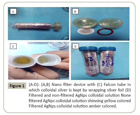
The procedure adopted for segregating bigger size particles from smaller size particles was to filter the bigger size silver particles colloidal silver with the help of a nano filter device. The device comprises of 2 knobs made up of hard plastic one washer and a fine filter with less than 100 nm pore size (Figure 1). The raw colloidal silver was freshly prepared and collected inside a 20 ml big syringe with silver foil wrapped and light protected. The shining yellow colored none filtered colloidal silver after filtration becomes amber color the shinning yellow color cause harm to eye ball. The amber colored solution was collected in a properly leveled falcon tube after filtration with silver foil wrapped and light and temperature protected as it was highly sensitive. Both non filtered and filtered colloidal silver bearing falcon tube should be properly leveled to differentiate between the two.
Transmission electron microscopy
To obtain the size and morphology of the Silver nano particles and to get the TEM image of the tissue which is 1, 1.5 and 10 lakh times magnified, TEM processing with viewing performed. TEM analysis of the PVP coated Sodium Borohydried stabilized Silver nano particles colloidal solution performed at an accelerating voltage of 100KV. Silver nano particles was examined in the form of colloidal solution suspended in NaCl (Ag-1.8mg/ml) and subsequently deposited on the form var/carbon coated TEM grid. Digital TEM camera was calibrated for size and measurement of the silver nano particular mass. Mean, Standard deviation, Individual differences from mean were calculated from procured information measuring over 100 Silver nano particles in the random fields of view with addition to it images procured by specific compacted cassettes which is viewed by special software named soft imaging viewer or SIS image viewer that showed general morphology of the scattered silver nano particles in the solution.
Transmission electron microscopy (TEM) analyses were performed on a FEI Tecnai G2 spirit twin transmission electron microscope equipped with Gatan digital CCD camera (Netherland) at 60 or 80 KV and to determine particle diameter using a Zeta Sizer-Nano ZS (Malvern Instruments, UK). (Zeta potential=-17.52 mV) The hydrodynamic diameter of AgNps was measured by dynamic light-scattering, and zeta potential was determined by laser Doppler micro electrophoresis.
Procedures for transmission electron microscopy of tissue
Cell suspensions of control and treated tissues were washed with 1XPBS (PH 7.2) and fixed in 2.5% Glutraldehyde prepared in Sodium Cacodylate (Ladd Research Industries, USA, Burlington) buffer (PH 7.2) for 2h at 40C. Cells were washed 3 times with 0.1M Sodium Cacodylate buffer and post fixed in 1% Osmium Tetroxide for 2 h. Fixed cells were washed with Sodium Cacodylate, dehydrated in acetone series (15-100%) and embedded in araldite-DDSA mixture (Ladd Research Industries, USA). After backing at 60°C, blocks were cut (60-80 nm thick) by an ultramicrotome (Leica EM UC7) and sections were stained by Uranyl acetate and Lead Citrate. Analysis of sections was done under FEI Tecnai G2 spirit twin transmission electron microscope equipped with Gatan digital CCD camera (Netherland) at 60 or 80 KV.
Confocal microscopy for assessment of apoptosis and necrosis
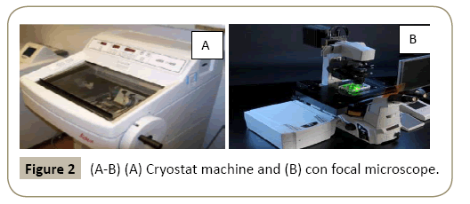
Procedure: For assessment of fresh tissue apoptosis and necrosis features visualization the of control and smaller size lower dose treated group (2.3-71 nm/0.5 mg and 1 mg /kg b.w.) and smaller size higher dose treated group (2.75-20 nm/10 and 15 mg/kg b.w.) liver tissue of fetuses collected after dissection and kept in Ependurf tube within 0.01% PBS solution and subjected to cryostat cutting, (Interdisciplinary School of Life Sciences, Banaras Hindu University under Lakhotiya Laboratory Department of Science (Department of Zoology and Botany)) slides are prepared and tissue sections were stained by Acridine Orange (Apoptosis), DAPI (Apoptosis) and Ethidium Bromide (1:250 dilution) (Necrosis) staining and a second wash with 0.01% PBS(Figure 2). The same day in which tissue dissected and slides prepared were exposed for con focal microscopy. Photograph taken by confocal microscope in coordinator of Interdisciplinary School of Life Sciences, Banaras Hindu University under Lakhotiya Laboratory Department of Science (Department of Zoology and Botany) and analyzed for cell apoptosis and necrosis assessment.
Procedure of cell grow for newly emerging and developing precursor hepatoblast and neuroblast stem cells of pregnant swiss albino mice and their fresh born fetuses: Newly emerging and developing precursor hepatoblast and neuroblast stem cells (After tissue crush with Mortar and Pestle isolated) of vital organs (Liver and Brain) of pregnant Swiss Albino mice and their fresh born fetuses were placed in flasks containing Dulbecco MEM nutrient media with 0.01% PBS (Phosphate Buffered Saline), L-glutamine and 1% antibiotic and were kept in a CO2 incubator conditioned with 4% CO2 at 37°C for 72 h. For harvesting cells, the cell culture medium was discharged, and the cells were treated with trypsin-EDTA (0.45 mL per flask). Cells were then transferred into 10 mL Eppendorf tubes and centrifuged at 5000 rpm for 5 min. The supernatant was discharged and the cells were used in the present study and further experimental studies.
Acridine orange and DAPI immunostaining assay for cytotoxicity: Newly emerging and developing precursor hepatoblast and neuroblast stem cells (After tissue crush with Mortar and Pestle isolated) of vital organs (Liver and Brain) of pregnant Swiss Albino mice and their fresh born fetuses (6 × 110 cells per well) were placed in 96-well plates containing Dulbecco MEM nutrient media with 0.01% PBS (Phosphate Buffered Saline), L-glutamine and 1% antibiotic. The plates were then kept in a CO2 incubator (37°C in 4% CO2) for 72 h, after which time, when the cells sticked to the base of the plate, the cell culture medium was replaced with fresh medium at different concentrations (30-150 μg/mL in aqueous suspensions) of colloidal small size AgNps were placed into the wells. Following incubation under the same conditions for an additional 72 h, Acridine Orange and DAPI immune staining reagent (2 μL) was added into each well. Upon incubation for an additional 8 h, the plates were immediately read in an Elisa Micro plate Reader (BioTek, USA) at 350 nm and 650 nm reference wavelengths.
Stem cell inflammation assay: Stem cell inflammation was determined using a con focal microscope view to monitor stem cell inflammation after treatment with colloidal small size AgNps in vitro newly emerging and developing precursor hepatoblast and neuroblast stem cells (After tissue crush with Mortar and Pestle isolated) of vital organs (Liver and Brain) of pregnant Swiss Albino mice and their fresh born fetuses (6 × 110 cells per well) were cultivated in Dulbecco MEM nutrient media with 0.01% PBS(Phosphate Buffered Saline), L-glutamine and 1% antibiotic, using E-Plate 96 (Roche) for cell-growth monitoring. The plates were kept in a CO2 incubator (37 °C in 4% CO2) during the experiment. At hour 27 of incubation, different concentrations of AgNps (30-150 μg/mL in medium) were added to the wells containing newly emerging and developing precursor hepatoblast and neuroblast stem cells (After tissue crush with Mortar and Pestle isolated) of vital organs (Liver and Brain) of pregnant Swiss Albino mice and their fresh born fetuses and cultivated under the same conditions mentioned above. For control groups, cell culture medium was added instead of colloidal small size AgNps solution. The attachment and logarithmic growth of the cells was measured every 15 min over 44 h.
Evaluation of apoptotic and necrotic stem cells: Immuno staining of Acridine Orange with DAPI and 1:250 dilutions Ethidium Bromide was performed on such tissue sections embedded stem cells to quantify and evaluate the number of apoptotic and necrotic cells in con focal microscope view on the basis of scoring cell nuclei. Newly emerging and developing precursor hepatoblast and neuroblast stem cells (After tissue crush with Mortar and Pestle isolated) of vital organs (Liver and Brain) of pregnant Swiss Albino mice and their fresh born fetuses (6 × 110 cells per well) were placed in 96 well plates containing Dulbecco MEM nutrient media with 0.01% PBS (Phosphate Buffered Saline), L-glutamine and 1% antibiotic at 37°C in a 4% CO2 humidified atmosphere in 96-well plates. Newly emerging and developing precursor tissue stem cells were treated with different concentrations of AgNps (30-150 μg/mL) for 72 h. The control group consisted of newly emerging and developing precursor hepatoblast and neuroblast stem cells treated with cell culture medium only. Both attached and detached cells were collected, then washed with 0.01% phosphate-buffered saline (PBS) and stained with Immuno staining of Acridine Orange with DAPI and 1:250 dilutions Ethidium Bromide solution for 1 to 2 min at 20°C temperature. Next, 10-20 μL of cell suspension was smeared onto a defrosted and autoclaved glass slide for examination by fluorescence and con focal microscope (Leica, Germany). With the Acridine Orange with DAPI, the nuclei of normal and treated cells were stained with green and blue fluorescence of high intensity, but apoptotic cells were stained a stronger orange or red fluorescence on con focal and fluorescence view. The apoptotic cells were mostly identified by morphological changes in the nucleus including nuclear fragmentation and chromatin condensation like mosaic presentation in a house floor [31,32]. Nuclei of necrotic cells were stained red by 1:250 dilutions Ethidium Bromide solution, as 1:250 dilutions Ethidium Bromide solution can cross the cell membrane of necrotic cells, which lack plasma membrane integrity. The 1:250 dilutions Ethidium Bromide solution cannot cross the non-necrotic cell membrane [33,34]. The numbers of apoptotic and necrotic cells were counted at 100 randomly chosen con focal microscopic fields using the 400X con focal fluorescent microscope objective. The number of apoptotic and necrotic cells were determined using a con focal and DSI259 Fluorescence Inverted Microscope (Leica, Germany) with Acridine Orange, DAPI and 1:250 dilutions Ethidium Bromide filters respectively. Data were expressed as a ratio of apoptotic or necrotic cells to normal cells.
Result
Characterization of small size silver nano particles
Optical properties of small size AgNps were determined by the Dynamic light scattering. The UV Vis absorption spectrum shows an intense absorption peak around 630 nm, originating from the surface Plasmon’ absorption of nano small sized AgNps. Under the conditions used for synthesis, silver nano particles had a relatively small size distribution around 2.75 nm to 20 nm, formed stable dispersions at the bottom of the colloid in Erlenmeyer’s flask, and were charged negatively. The zeta potential value means the rate of electrical conduction in the solution matrix was found -17.52 mV in Auto Correlation Function Graph with mobility distribution and Equation of state plot, which is an optic interference property of sodium borohydride stable colloids. In the zeta analysis, the suspension of silver nano particles consisted of particles with an average size of 11.25 nm in hydrodynamic diameter. Most of the silver nano particles ranged from 2.75 to 20 nm in size. The average size of the particles on the substrates was 11.25 nm with a standard deviation of 1.2 nm.
Cytotoxicity of newly emerging and developing precursor hepatoblast and neuroblast stem cells of pregnant Swiss Albino mice and their fresh born fetuses exposed to small size silver nano particles had alterations in cell shape and morphology, whereas control cells demonstrated no change in morphology. According to Acridine Orange and DAPI assay results, small size silver nanoparticle concentrations significantly affected their impact on cell viability in a concentration and dose-dependent manner. The lowest mortality rate was obtained at a silver nanoparticle concentration of 30 μg/mL, whereas the highest mortality rate was obtained at 150 μg/mL, which was the highest concentration tested. Small size AgNps were cytotoxic on newly emerging and developing precursor hepatoblast and neuroblast stem cells of pregnant Swiss Albino mice and their fresh born fetuses with a half-maximal inhibiting concentration value (LC50) of 30 μg/mL.
Cell inflammation effect of small size silver nano particles
Inflammation effects of small size silver nano particles were evaluated on newly emerging and developing precursor hepatoblast and neuroblast stem cells of pregnant Swiss Albino mice and their fresh born fetuses based on the Con focal microscopy view which was found concentration and dosedependent.
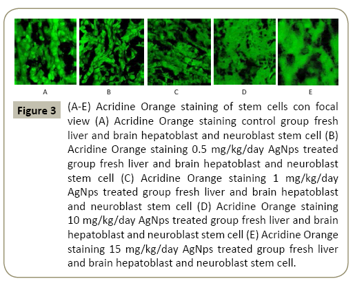
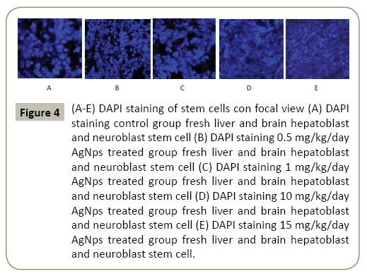
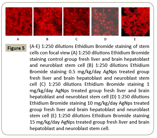
Estimation of live and dead cells and their fate of individual staining of 2.75 to 20 nm mean size AgNps treated group liver and brain tissue of fetuses (Figures 3-5).
div class="well well-sm">Figure 3: (A-E) Acridine Orange staining of stem cells con focal view (A) Acridine Orange staining control group fresh liver and brain hepatoblast and neuroblast stem cell (B) Acridine Orange staining 0.5 mg/kg/day AgNps treated group fresh liver and brain hepatoblast and neuroblast stem cell (C) Acridine Orange staining 1 mg/kg/day AgNps treated group fresh liver and brain hepatoblast and neuroblast stem cell (D) Acridine Orange staining 10 mg/kg/day AgNps treated group fresh liver and brain hepatoblast and neuroblast stem cell (E) Acridine Orange staining 15 mg/kg/day AgNps treated group fresh liver and brain hepatoblast and neuroblast stem cell.
Figure 4: (A-E) DAPI staining of stem cells con focal view (A) DAPI staining control group fresh liver and brain hepatoblast and neuroblast stem cell (B) DAPI staining 0.5 mg/kg/day AgNps treated group fresh liver and brain hepatoblast and neuroblast stem cell (C) DAPI staining 1 mg/kg/day AgNps treated group fresh liver and brain hepatoblast and neuroblast stem cell (D) DAPI staining 10 mg/kg/day AgNps treated group fresh liver and brain hepatoblast and neuroblast stem cell (E) DAPI staining 15 mg/kg/day AgNps treated group fresh liver and brain hepatoblast and neuroblast stem cell.
Figure 5: (A-E) 1:250 dilutions Ethidium Bromide staining of stem cells con focal view (A) 1:250 dilutions Ethidium Bromide staining control group fresh liver and brain hepatoblast and neuroblast stem cell (B) 1:250 dilutions Ethidium Bromide staining 0.5 mg/kg/day AgNps treated group fresh liver and brain hepatoblast and neuroblast stem cell (C) 1:250 dilutions Ethidium Bromide staining 1 mg/kg/day AgNps treated group fresh liver and brain hepatoblast and neuroblast stem cell (D) 1:250 dilutions Ethidium Bromide staining 10 mg/kg/day AgNps treated group fresh liver and brain hepatoblast and neuroblast stem cell (E) 1:250 dilutions Ethidium Bromide staining 15 mg/kg/day AgNps treated group fresh liver and brain hepatoblast and neuroblast stem cell.
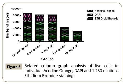
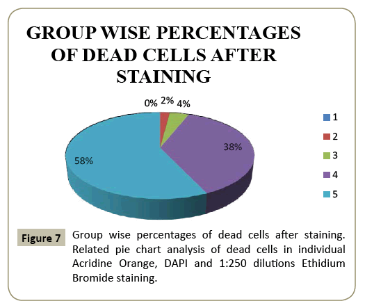
Image-j estimation of apoptotic and necrotic cell (control group and 0.5, 1, 10, 15 mg/kg/day 2.75 to 20nm mean small size AgNps treated groups apoptotic and necrotic cell estimation from fresh tissue from control group and treated groups and evaluation of the fate of live and dead cells (Tables 1-3) (Figures 6 and 7).
Figure 6: Related column graph analysis of live cells in individual Acridine Orange, DAPI and 1:250 dilutions Ethidium Bromide staining.
Table 1 Live cell estimation of Acridine Orange stained fresh hepatoblast and neuroblast stem cells.
| Slice | Count | Total area | Average size | Mean | Mode |
|---|---|---|---|---|---|
| Ctrl | 6254 | 246568.0 | 1887.446 | 64.463 | 58.807(AO) |
| 0.5 | 6180 | 237699.0 | 1821.876 | 62.456 | 57.962(AO) |
| 1 | 6172 | 224759.0 | 1799.776 | 60.356 | 56.862(AO) |
| 10 | 3988 | 161303.0 | 1707.720 | 54.450 | 31.473(AO) |
| 15 | 2983 | 161003.0 | 1697.720 | 49.450 | 26.473(AO) |
Table 2 Live cell estimation of DAPI stained fresh hepatoblast and neuroblast stem cells.
| Slice | Count | Total area | Average size | Mean | Mode |
|---|---|---|---|---|---|
| Ctrl | 2311 | 71888.00 | 71.107 | 56.876 | 254.811(DAPI) |
| 0.5 | 1885 | 67169.00 | 70.084 | 55.803 | 249.094(DAPI) |
| 1 | 1875 | 67069.00 | 69.984 | 54.703 | 248.994(DAPI) |
| 10 | 300 | 11612.00 | 8.127 | 36.500 | 231.777(DAPI) |
| 15 | 175 | 11212.00 | 5.127 | 31.500 | 226.777(DAPI) |
Table 3 Live cell estimation of 1:250 dilutions Ethidium Bromide stained fresh hepatoblast and neuroblast stem cells.
| Slice | Count | Total area | Average size | Mean | Mode |
|---|---|---|---|---|---|
| Ctrl | 96 | 4097 | 10.42677 | 4.0392 | 5.7618(EB) |
| 0.5 | 93 | 4722 | 9.21742 | 3.7453 | 4.0430(EB) |
| 1 | 83 | 4688 | 9.18742 | 3.6453 | 4.0403(EB) |
| 10 | 23 | 1973 | 8.62286 | 3.2392 | 3.4888(EB) |
| 15 | 18 | 1968 | 7.91686 | 3.0392 | 2.9888(EB) |
During the study we observed that the number of live stem cell after Acridine orange, DAPI and 1:250 dilutions Ethidium Bromide staining in 0.5, 1, 10 and 15mg/kg b.w. 2.75 to 20 nm mean size range AgNps oral gavages treated group was less than control. AO, DAPI and 1:250 dilutions Ethidium Bromide is used to visualize nuclear morphology hence less number of live stem cells in 0.5, 1, 10 and 15 mg/kg b.w. small size AgNps oral gavages treated group determine that in 0.5, 1, 10 & 15 mg/kg b.w. small size AgNps oral gavages treated group affected mice more number of cell must have undergone apoptosis and necrosis leading to fragmentation of cell which does not occur in normal cell. So a conclusion comes out of it. In small size AgNps colloidal solution repeated oral gavages testing experimentation on hepatoblast and neuroblast stem cells of pregnant Swiss albino mice and their fetuses shows rational effects of smaller size silver particles on tissue (liver and brain tissue) level of fetuses, smaller particles shows more injury and higher dose shows more injury on cellular organelles including death, degeneration and fragmentation. There is a rational effect of apoptosis and necrosis. The live cell estimation shows chronological decrease of number as dose of small size silver nano particles increases the dead cell estimation shows chronological increase of percentages as dose of small size silver nano particles increases(above pie chart analysis). The above analysis proves a definite feature of stem cell cytotoxicity response after repeated oral gavages (5 to 15 gestational age of mother) small size AgNps application on hepatoblast and neuroblast stem cells of Pregnant Swiss albino mice and their fetuses which is purely concentration and dose dependent [35].
Discussion
Small size silver nano particles are metallic nano particles with highly utilized physiochemical and surface properties and have been utilized for various biomedical purposes, such as cancer diagnostic, wound dressings medication cream, jewelry and cosmetics, and in the medical industry as prosthetics-rust coating agents [36,37]. Anyway, most of the nano experimental researches in the past demonstrated small size nano silver (AgNps) may induce genotoxicity and cytotoxicity (Apoptosis and necrosis) in carcinomatous and for experimental purpose available normal cell tracts [38]. In this present study, market available synthesized small size AgNps were also determined to be apoptotic, necrotic and cytotoxic to certain phase on newly emerging and developing hepatoblastoma and neuroblastoma stem cells of primate vertebrate and their fresh born fetuses with an LC 50 value of 30 μg/mL. Very importantly, the mode of apoptogenic and necrogenic sequence with cytotoxic consequences may vary at different level of concentrations, as the apoptotic and necrotic indexes increased depending on the small size AgNps concentration. However, at higher concentrations of small size nano silver the necrotic effects became more dominant corresponding to dose, with a moderate number of apoptotic and cytotoxic consequences corresponding to same dose. A famous international nano scientist also in past erased an apoptotic , necrotic and cytotoxic effects on newly emerging and developing hepatoblastoma and neuroblastoma stem cells of primate vertebrate and their fresh born fetuses in vitro induced by small size AgNps used at low and moderate concentrations [39]. Small size AgNps induce toxic cell responses often specific to culture cell types, resulting in varying degrees of apoptosis, necrosis and cytotoxicity. Based on silver nanoparticle length, breadth of morphology, concentration, and application time, these small size silver nano particles exhibits various levels of apoptosis, necrosis & cytotoxicity in vitro in hepatoblastoma and neuroblastoma stem cells and other skin and lung stem cells [40-42]. One of the world famous nano scientist showed that small size AgNps at a lower most concentration (10-37 μg/mL) induces a depletion in chondriblastic activity. More or less same type of results have been reported in homo sapiens (0.6-4 μg/ mL) and rodent (15-55 μg/mL) hepar cells, lung alveolar macrophages (15-80 μg/mL), mouse skin fibroblastoma stem cells and hepatoblast stem cells (40 -50μg/mL), and mouse germ line stem cells (5 -20 μg/mL) [43,44]. The rate of apoptosis, necrosis and cytotoxicity increased with the depletion of the silver nanoparticle size and increase of surface area. Furthermore, the cells tested by Liu scientist and groups showed varied in apoptosis, necrosis and cytotoxicity rate [45,46]. For an instance, the LC 50 value of AgNps of 2.75 to 20 nm in size range in hepatoblast stem cells was 13.33 ± 0.361 μg/mL, while it was 45.96 ± 4.75 μg/mL in neuroblast stem cells. In our study, the size of AgNps varied from 2.75 nm to 20 nm as determined by TEM and spectroscopic studies. Using an Immunostaining cytotoxicity test, we found that the LC 50 value was 30 μg/mL for our experiment applied small size AgNps. We determined apoptotic, necrotic and cytotoxicity effects induced by our experiment applied small size AgNps on hepatoblast and neuroblast stem cells of pregnant Swiss Albino mice and their fresh born fetuses even at a concentration as low as 30 μg/mL. Furthermore, the inflammation of hepatoblast and neuroblast stem cells of pregnant Swiss Albino mice and their fresh born fetuses was interfered with small size our experiment applied AgNps in a dose and concentration-dependent manner, starting at a concentration as low as 30 μg/mL up to highest 150 μg/mL. We think that the data generated through this study will be useful for our proposed future studies, in which we will focus on mitochondrial autophagy on hepatoblast and neuroblast stem cells of pregnant Swiss Albino mice and their fresh born fetuses using small size fresh made colloidal AgNps solution. The mechanism of cytotoxicity induced by small size our experiment applied AgNps is also of scientific interest to other researchers. Small size AgNps are reported to induce severe cellular structural and histomorphological damage, accumulate in chondroblast, and contribute to singlet oxygen misbalance load [47]. A very internationally popular nano scientist discovered that small size AgNps at a concentration of 10-37 μg/mL caused depletion in chondroblastic activity. Similar apoptotic, necrotic and cytotoxicity results have been reported in homosapien and rodent hepatoblast stem cells, lung alveolar macrophages, mouse skin fibroblast stem cells and hepatoblast stem cells, and mouse germ line stem cells. With addition to this quote, another famous nano scientist reported that small size AgNps principally deviates GSSH levels and increased generation of singlet oxygen genome (ROS) in cells of the body [48]. It is well known and an internationally established scientific fact that singlet oxygen genome are highly reactive and cause oxidative harm to deoxy ribo nucleic and other cell enzymes [49,50]. Generation of excessive intracellular singlet oxygen genome leads to apoptosis and necrosis [51]. Evidenced by the fact that an increase in singlet oxygen genome levels is correlated with maximum deoxy ribo nucleic acid breakage and high levels of apoptosis and necrosis. Anyway, the cytotoxicity effect may be halfly due to direct action of Silver+ ions released from small size our experiment applied nano silver. 2 internationally famous Indian Scientist reported that small size AgNps were up taken by macrophages via receptormediated phagocytosis, and Silver+ ions were released from our experiment applied nano silver [52]. The free Silver+ ions in due course enter in various intra cytoplasm and nucleolus structures and tracts, including chondroblastic activities inducing stress pathways and arises apoptosis. Thus, future researches are required to discover the remain hidden fact that the small size AgNps-induces more apoptosis and necrosis rate in comparison to bigger size from the retrospective action of singlet oxygen genome. In the present research study, we preferred to synthesize small size AgNps of 2.75 to 20 nm range and 71.5 nm in hydrodynamic diameter, which is a considerably smaller size for testing cytotoxicity. As past research reported by world famous Southeast Asian nano scientist, that nano small sized silver are more toxic than bigger size and micro sized nano silver particles [53]. With assimilation to the existing, the small size nano silver that we synthesized in this experimental study are quite cytotoxic despite their negative zeta potential charge (-17.52 mV). A change in morphology of the hepatoblast and neuroblast stem cells was observed upon small size AgNps treatment as the first indication of apoptosis, necrosis and cytotoxicity. Predominant depletion in hepatoblast and neuroblast stem cell viability was observed after experiment, probably as a result of reduction in ATP production, generation of singlet oxygen genome (ROS), and damage to the chondroblastic respiratory chain activity. The singlet oxygen genome over secretion is main cause for triggering DNA damage, followed by cell cycle arrest (Programmed cell death) at G2/M stage. The cells arresting (Programmed cell death) at G2/M stage are not undergoing massive apoptosis or necrosis, and no fragmented nuclei or necrotic hepatoblast and neuroblast stem cells were observed. Hence we speculate that the accumulation of hepatoblast and neuroblast stem cells at the G2/M interface is associated with DNA repair which could lead to programmed cell death or survival at a later stage. All the data taken together suggest that smaller size silver nanoparticles at the 2.75 to 20 nm range and 30 μg/mL concentrations used resulted in G2/M arrest in the hepatoblast and neuroblast stem cells, which might lead to programmed cell death if repair pathways were unsuccessful.
On examination of con focal microscopy images of control, 0.5 and 1 mg/kg/day treated group stained by Acridine orange, DAPI and 1:250 dilutions Ethidium Bromide stain for apoptosis and necrosis showed dull green and blue appearance for apoptotic presentation and dazzling orange read appearance for normal and dull orange red for necrotic tissue. The apoptotic tissues were found surrounding normal tissue like puff of powder thrown on floor. The necrotic tissues were found in cluster like small piece of stone with powder mix thrown on floor looks like intricate network. The intensity of such sign and symptoms were found milder in lower dose group whereas in 10 and 15 mg treated higher dose group same sign and symptoms were seen but the intensity of it was found higher. So 0.5 1 mg/kg/day can be comparable with 10 and 15 mg/kg/day. On thorough evaluation of fate of cells after counting live cells and dead cells following use of Acridine Orange, DAPI and 1:250 dilutions Ethidium Bromide staining technique our study come to a conclusion that number of live cells decrease with increase dose of small size colloidal fresh made silver nano particles whereas percentages of dead cells increase with increase dose of same colloidal fresh made silver nano particles after repeated oral gavages testing on animals. Our study findings shows matching with study findings of Chakraborty et al. [54], Langheinrich et al. [55], for apoptosis and Sheng et al. [56] for necrosis Asharani et al. [15], Hackenberg et al. [51] Foldbjerg et al. [19] Singh and Ramarao [52] for apoptosis and necrosis [54-56]. The results of the study done by a super special worldwide recognized nano scientist indicated that small size silver nanoparticle activated stress-responsive gene GSS and immune reaction genes CXCL13 and MARCO. Small size silver nanoparticle induces inflammatory cytokine TNF-α release, active singlet oxygen genome and endoplasmic reticulum (ER) stress response in zebra fish liver cells [57]. Another eminent worldwide recognized Chinese nano scientist found small size nano silver-treated hepatoblast stem cells have the up-regulated gene expression of CXCL13 and MARCO to induce apoptosis and inflammation which matches with the present study report [58].
Conclusion
In conclusion, small size AgNps of 2.75 to 20 nm size and 71.5 nm in hydrodynamic diameter and with a zeta potential of -17.52 mV express apoptosis, necrosis and cytotoxicity on the hepatoblast and neuroblast stem cells with an LC50 value of 30-150 μg/mL. Thus, they can be used in vitro at concentrations of up to 150 μg/mL. Small size silver nanoparticles in fresh made colloidal form induce apoptosis and necrosis at lower concentrations, but induce necrosis only at higher concentrations in pregnant Swiss Albino mice and fetuses isolated and processed hepatoblast and neuroblast stem cells. Further researches are required to reopen and discover the pathway of the apoptotic and necrotic effects of small size silver nanoparticles.
References
- Oberdorster G, Oberdorster E, Oberdorster J (2005) Nano toxicology: an emerging discipline evolving from studies of ultrafine particles. Environ Health Perspect 113: 823-839.
- Tan WB, Jiang S, Zhang Y (2007) Quantum-dot based Nano particles for targeted silencing of HER2/neu gene via RNA interference. Biomaterials 28: 1565-1571.
- Yoon K, Hoon B, Park JH, Hwang J (2007) Susceptibility constants of Escherichia coli and Bacillus subtilis to silver and copper nanoparticles. Sci Total Environ 373: 572-575.
- Kreuter J, Gelperina S (2008) Use of nanoparticles for cerebral cancer. Tumori 94: 271-277.
- Su J, Zhang J, Liu L, Huang Y (2008) Exploring feasibility of multicolored CdTe quantum dots for in vitro and in vivo fluorescent imaging. J Nanosci Nanotechnol 8: 1174-1177.
- Hackenberg S, Scherzed A, Kessler M (2010) Zinc oxide nanoparticles induce photocatalytic cell death in human head and neck squamous cell carcinoma cell lines in vitro. Int J Oncol 37: 1583-1590.
- Cho K, Park J, Osaka T (2005) Te study of antimicrobial activity and preservative effects of nano silver ingredients. Electrochim Acta 51: 956-960.
- Kim J, Kuk E, Yuk N (2006) Mode of antibacterial action of silver nanoparticles. J Proteome Res 5: 916-924.
- Ahmed M, Karns M, Goodson M (2008) DNA damage response to different surface chemistry of silver nanoparticles in mammalian cells. Toxicol Appl Pharmacol 233: 404-410.
- Johnston HJ, Hutchison G, Christensen FM (2010) A review of the in vivo and in vitro toxicity of silver and gold particulates: particle attributes and biological mechanisms responsible for the observed toxicity. Crit Rev Toxicol 40: 328-346.
- Zanette C, Pelin M, Crosera M (2011) Silver nanoparticles exert a long-lasting anti-proliferative effect on human keratinocyte HaCaT cell line. Toxicol in Vitro 5: 1053-1060.
- Chen X, Schluesener HJ (2008) Nano silver: a Nano product in medical application. Toxicol Lett 176: 1-12.
- Chaloupka K, Malam Y, Seifalian AM (2010) Nano silver as a new generation of Nano product in biomedical applications. Trends Biotechnol 28: 580-588.
- Yoon K, Hoon B, Park JH (2007) Susceptibility constants of Escherichia coli and Bacillus subtilis to silver and copper nanoparticles. Sci Total Environ 373: 572-575.
- Asharani PV, Low Kah Mun G, Hande MP (2009) Cytotoxicity and genotoxicity of silver nanoparticles in human cells. ACS Nano 3: 279-290.
- Hussain SM, Hess KL, Gearhart JM (2005) In vitro toxicity of nanoparticles in BRL 3A rat liver cells. Toxicol. In-Vitro 1: 975-983.
- Kim YS, Song MY, Park JD (2010) Sub-chronic oral toxicity of silver nanoparticles. Part Fibre Toxicol 7: 20-31.
- Sonoda E, Sasaki MS, Buerstedde JM (1998) Rad51-defcient vertebrate cells accumulate chromosomal breaks prior to cell death. EMBO J 15: 598-608.
- Foldbjerg R, Olesen P, Hougaard M (2009) PVP-coated silver nanoparticles and silver ions induce reactive oxygen species, apoptosis, and necrosis in THP-1 monocytes. Toxicol Lett 190: 156-162.
- Hsin Y, Chen C, Huang S (2008) Te apoptotic effect of Nano silver is mediated by ROS- and JNK-dependent mechanism involving the mitochondrial pathway in NIH3T3 cells. Toxicol Lett 179: 130-139.
- Hess R, Jones L, Schlager JJ (2008) unique cellular interaction of silver nanoparticles: size-dependent generation of reactive oxygen species. J Phys Chem B 112: 13608-13619.
- Li GY, Osborne NN (2008) Oxidative-induced apoptosis to an immortalized ganglion cell line is caspase independent but involves the activation of poly (ADP-ribose) polymerase and apoptosis-inducing factor. Brain Res 188: 35-43.
- Liu W, Wu Y, Wang C (2010) Impact of silver nanoparticles on human cells: effect of particle size. Nano toxicology 4: 319-330.
- Liu X, Lee PY, Ho CM (2010) Silver nanoparticles mediate differential responses in keratinocytes and fibroblasts during skin wound healing. Chem Med Chem 1: 468-475.
- Sur I, Altunbek M, Kahraman M (2012) The influence of the surface chemistry of silver nanoparticles on cell death. Nanotechnology 23: 375102.
- Oberley T, Sioutas C, Yeh JI (2006) Comparison of the abilities of ambient and manufactured nanoparticles to induce cellular toxicity according to an oxidative stress paradigm. Nano Lett 6: 1794-1807.
- Solomon SD, Bahadory M, JeyarajasingamAV (2007)Journal of Chemical Education84: 322-325.
- Berne BJ, Pecora R (2010) Dynamic Light Scattering. Courier Dover Publications.
- Schneider CA, Rasband WS, Eliceiri KW (2012) NIH Image to Image-J: 25 years of image analysis. Nat Methods 9: 671-675.
- Reimer L (1997) Transmission Electron Microscopy: Image Formation and Microanalysis 4th Edn. Springer.
- Van England M, Nieland LJ, Ramaekers FC (1998) Annexin V-afnity assay: a review on an apoptosis detection system based on phosphatidylserine exposure. Cytometry 31: 1-9.
- Turk M, Rzaev ZMO, Kurucu G (2010) Bioengineering functional copolymers. XII. Interaction of boron-containing and PEO branched derivatives of poly (MA-alt-MVE) with HeLa cells. Health 2: 51-61.
- Kamphaus GD, Colorado PC, Panka DJ (2000) Canstatin, a novel matrix-derived inhibitor of angiogenesis and tumor growth. J Biol Chem 275: 1209-1215.
- Tice RR, Agurell E, Anderson D (2000) Single cell gel/comet assay: guidelines for in vitro and in vivo genetic toxicology testing. Environ Mol Mutagen 35: 206-221.
- Cho KS, Lee EH, Choi JS, Joo CK (1999) Reactive oxygen species-induced apoptosis and necrosis in bovine corneal endothelial cells. Investigative Ophthalmology & Visual Science 40: 911-919.
- Tian J, Wong KK, Ho CM (2007) Topical delivery of silver nanoparticles promotes wound healing. Chem Med Chem 2: 129-136.
- Wijnhoven SWP, Peijnenburg WJGM, Herberts CA (2009) Nano-silver - a review of available data and knowledge gaps in human and environmental risk assessment. Nano toxicology 3: 109-138.
- Yoon K, Hoon B, Park JH, Hwang J (2007) Susceptibility constants of Escherichia coli and Bacillus subtilis to silver and copper Nano particles. Sci Total Environ 373: 572-575.
- Gopinath P, Gogoi SK, Chattopadhyay A (2008) Implications of silver nanoparticle induced cell apoptosis for in vitro gene therapy. Nanotechnology 19: 075104.
- Pan Y, Neuss S, Leifert A (2007) Size-dependent cytotoxicity of gold nanoparticles. Small 3: 1941-1949.
- Park S, Lee YK, Jung M (2007) Cellular toxicity of various inhalable metal nanoparticles on human alveolar epithelial cells. Inhal Toxicol 19: 59-65.
- Hess R, Jones L, Schlager JJ (2008) unique cellular interaction of silver nanoparticles: size-dependent generation of reactive oxygen species. J Phys Chem B 112: 13608-13619.
- Braydich-Stollen L, Hussain S, Schrand A (2005) In vitro cytotoxicity of nanoparticles in mammalian germline stem cells. Toxicol Sci 88: 412-419.
- Liu W, Wu Y, Wang C (2010) Impact of silver nanoparticles on human cells: effect of particle size. Nanotoxicology 4: 319-330.
- Liu X, Lee PY, Ho CM (2010) Silver nanoparticles mediate differential responses in keratinocytes and fbroblasts during skin wound healing. Chem Med Chem 1: 468-475.
- Mahmood M, Casciano DA, Mocan T (2010) Cytotoxicity and biological effects of functional nanomaterials delivered to various cell lines. J Appl Toxicol 30: 74-83.
- Asharani PV, Hande MP, Valiyaveettil S (2009) Anti-proliferative activity of silver nano particles. BMC Cell Biol 10: 65-79.
- Oberley T, Sioutas C, Yeh JI (2006) Comparison of the abilities of ambient and manufactured nano particles to induce cellular toxicity according to an oxidative stress paradigm. Nano Lett 6: 1794-1807.
- Turrens J (2003) mitochondrial formation of reactive oxygen species. J Physiol 552: 335-344.
- Boonstra J, Post JA (2004) Molecular events associated with reactive oxygen species and cell cycle progression in mammalian cells. Gene 337: 1-13.
- Hackenberg S, Scherzed A, Kessler M (2011) Silver nanoparticles: evaluation of DNA damage, toxicity, and functional impairment in human mesenchymal stem cells. Toxicol Lett 201: 27-33.
- Singh RP, Ramarao P (2012) Cellular uptake, intracellular trafficking, and cytotoxicity of silver nanoparticles. Toxicol Lett 213: 249-259.
- Sohaebuddin SK, Tevenot PT, Baker D (2010) Nano material cytotoxicity is composition, size, and cell type dependent. Part Fibre Toxicol 7: 22-44.
- Chakraborty A, Uechi T, Higa S (2009) Losso fribosomal protein L11 affects zebra fish embryonic development through a p53-dependent apoptotic response. PLoS ONE 4: e4152.
- Langheinrich U, Hennen E, Stott G (2002) Zebra fish as a model organism for the identification and characterization of drugs and genes affecting p53signaling. Curr Biol 12: 2023-2028.
- Sheng G, Luo K, Zheng J (2008) enzymatically induced formation of neodymium hex cyanoferrate nanoparticles on the glucose oxidase/chitosan modified glass carbon electrode for the detection of glucose. Biosensors and Bioelectronics 24: 429-434.
- Christen V, Capelle M, Fent K (2013) Silver nanoparticles induce endoplasmic reticulum stress response in zebra fish Toxicol. Appl Pharmacol 272: 519-528.
- Cha HW, Hong YG, Choi MJ (2008) Comparison of acute responses of mice livers to short term exposure to Nano-sized or micro-sized silver particles. Biotechnol Lett 30: 1893-1899.
Open Access Journals
- Aquaculture & Veterinary Science
- Chemistry & Chemical Sciences
- Clinical Sciences
- Engineering
- General Science
- Genetics & Molecular Biology
- Health Care & Nursing
- Immunology & Microbiology
- Materials Science
- Mathematics & Physics
- Medical Sciences
- Neurology & Psychiatry
- Oncology & Cancer Science
- Pharmaceutical Sciences
