Antifungal Efficacy of Silver Nanoparticles against Plant Pathogenic Fungi
Nitaswini Dutta1, Shrayana Ghosh1, Madhura Das1, Surekha Kundu2 and Sarmistha Ray1*
1Department of Biotechnology, Amity University Kolkata, Kolkata, India
2Department of Botany, University of Calcutta, Kolkata, India
- *Corresponding Author:
- Sarmistha Ray
Department of Biotechnology,
Amity University Kolkata, Kolkata,
India,
E-mail: sarmiray86@gmail.com
Received date: March 03, 2023, Manuscript No. IPRJPP-23-16008; Editor assigned date: March 06, 2023, PreQC No. IPRJPP-23-16008 (PQ); Reviewed date: March 17, 2023, QC No. IPRJPP-23-16008; Revised date: March 27, 2023, Manuscript No. IPRJPP-23-16008 (R); Published date: April 03, 2023, DOI: 10.36648/ iprjpp.6.1.160
Citation: Dutta N, Ghosh S, Das M, Kundu S, Ray S (2023) Antifungal Efficacy of Silver Nanoparticles against Plant Pathogenic Fungi. Res J Plant Pathol Vol.6 No.1:160.
Abstract
The given finding is concerned with the fungicidal properties of silver nanoparticles used for antifungal treatment against plant pathogen. We have used biosynthesized silver nanoparticles (TcAgNp) at various concentrations 10%, 20%, 50% respectively. The plant pathogenic fungi were treated with the given concentrations on Malt Extract Agar (MEA). We calculated fungal inhibition both on plate measuring the radial diameter and also % spore germination inhibition in order to evaluate the antifungal efficacy. The result exhibited that AgNps possesses antifungal properties. At maximum concentration (50%) of silver nanoparticles it showed significant inhibition of plant pathogenic fungi that was observed on MEA. Our results open new avenues for the potential of biosynthesized silver nanoparticles to effectively control this fungal pathogen and set the basis for establishing safe strategy in its management programs in India.
Keywords
Nanofungicides; Silver nanoparticles; Antifungal; Efficacy; Spore germination frequency
Introduction
Plant diseases have greatly affected the agricultural production rate so various natural and artificial methods are employed for crop protection. Among various disease control methods use of artificial pesticides is the most prevalent. But the excessive use of these pesticides have resulted in contamination in water resources and food products affecting food chain [1]. Nanofungicides have added advantages over chemical pesticides as they are encapsulated by synthetic or natural protein coat, provides stability and its water solubility provides easier uptake through soil percolations without being readily dispersed in the atmosphere causing less environmental contamination it also reduces the irritation of the human mucous-membrane, the phytotoxicity and the environmental damage to other untargeted organisms and even to the crops [2]. One of the most potential eco-friendly method is the ‘green synthesis’ of nanoparticles. Among various nanoparticles used as nanoagrochemicals but very few reports are there that shows the importance of silver nanoparticles in management of plant diseases [3].
Silver plays an inhibitory roles and biological agents against microorganisms [4]. The antifungal activities of silver nanoparticles increase the efficacy of disease suppression. This nanosilver provides inhibition against reported certain reported harmful fungal pathogens Penicillium sp., Aspergillus sp. [5].
The objectives of the present study were to determine the inhibitory properties of silver nanoparticles against various commercially important plant pathogenic fungi and to evaluate the efficacy of silver compounds for suppression of plant pathogenic fungi in vitro.
Materials and Methods
Silver nanoparticles (TcAgNp) used for the following experiment
The silver nanoparticles were synthesized from Truncatoflabellum crassum (TcAgNp) and have been prepared using standard, published protocol using cell filtrate by extracellular synthesis of nanoparticles [6]. Quantitation of silver nanoparticles showed the concentration to be 28 mg/L.
Fungi and growth media
The selected fungal pathogen Magnaporthe oryzae used as it causes devastating rice blast disease along with early blight disease caused by Alternaria solani. The fungi were grown on Malt Extract Agar (MEA) in order to evaluate the antifungal inhibitory activities of silver nanoparticles in culture media.
In vitro assay
In vitro assay For the in vitro assay experiment was performed on Malt Extract Agar (MEA) treated with different concentrations (i.e. 10%, 20%, 50%) of silver nanoparticles. Two ml of AgNps having different concentrations was added into growth media prior to plating in a petri dish (90 mm x 15 mm). The media containing silver nanoparticles was incubated at room temperature. The observations were made after 24 hrs and 96 hrs of incubation, agar plugs of uniform size (diameter, 4 mm) containing fungi were inoculated simultaneously at centre of each petri dish containing silver nanoparticles.
Data analysis
radial growth of fungus was observed and recorded. The rate of radial inhibition (%) was calculated using the given formula (R-r)/R x 100 where R is the radial growth of mycelia for control plate and r is the radial growth of fungal mycelia on the treated plate with silver nanoparticles.
Effect of nanopart cles on the spore viability of plant pathogenic fungus Magnaporthe oryzae and Alternaria solani
The plant pathogenic fungus Magnaporthe oryzae was used to study the effect of silver nanoparticles on the rate of its spore germination. Spores were scraped from a 10 day old Magnaporthe oryzae and Alternaria solani culture grown on PDA plate. Spore suspension was made in sterile distilled water at a concentration of 1 x 10⁵ with the help of a haemocytometer. Silver nanoparticles were made of different concentrations such as 0.28, 0.56, 1.4 μg of nanoparticles in 100 μl solution. Equal volumes of diluted nanoparticles suspensions were added to 50 μl of Alternaria solani and Magnaporthe oryzae spore suspension. The culture tubes were incubated at 28°C. The spores were observed at 0 hrs, 2 hrs, 4 hrs, 6 hrs with lactophenol solution under a compound light microscope. The spore germination frequency and germ tube length were measured for different concentrations [6].
Result and Discussion
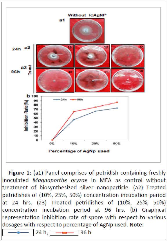
The inhibition effect of AgNPs at different concentrations was analysed in MEA. For in vitro assays exhibited higher inhibition of fungal growth were recorded at a concentration of 50% both for 24 hrs and 96 hrs time duration after post innoculation. In addition, specifically the rice blast fungi showed growth inhibition with the increment of incubation time. The lowest level of inhibition was observed against Magnaporthe oryzae on Malt Extract Agar (MEA) treated with a 10% concentration of AgNPs. It has been noted that with increase in time period that is at 96 hrs it exhibited highest inhibition rate of fungal growth in comparison to 24 hrs. That confers the stability of silver nanoparticle ever after 4 days (Figures 1a and 1b).
Figure 1: (a1) Panel comprises of petridish containing freshly inoculated Magnaporthe oryzae in MEA as control without treatment of biosynthesized silver nanoparticle. (a2) Treated petridishes of (10%, 25%, 50%) concentration incubation period at 24 hrs. (a3) Treated petridishes of (10%, 25%, 50%) concentration incubation period at 96 hrs. (b) Graphical representation inhibition rate of spore with respect to various dosages with respect to percentage of AgNp used. Note:
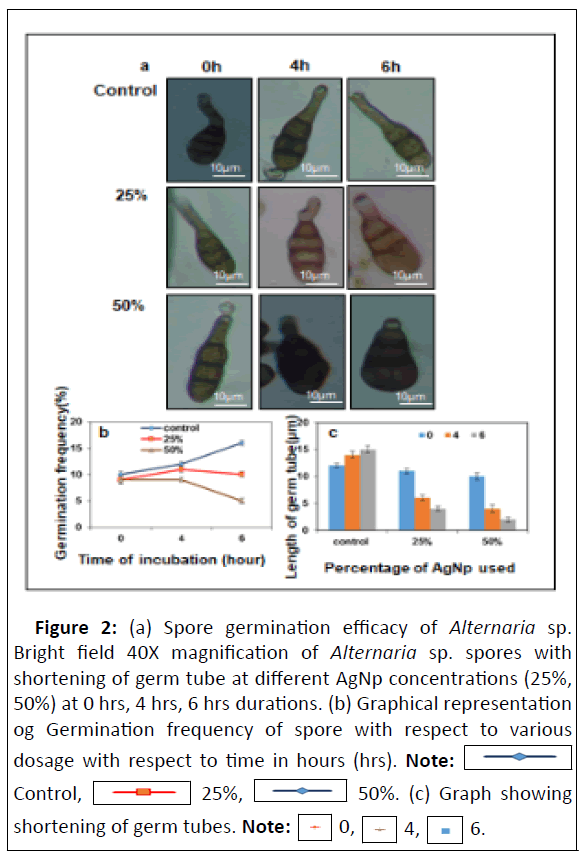
For spore germination assay, the germination frequency was seen to be inhibited at 50% concentration of silver nanoparticles. With progress in time starting from 0 hrs to 6 hrs the germination frequency drastically decreased to 10%. Even the germ tube length decreases with 50% concentration of silver nanoparticles, this is in accordance with the germination frequency assay for Magnaporthe oryzae that is with increase of time there is decrease in germ length. Similar results were observed with early blight pathogen Alternaria solani. The observation recorded may be due to the reports as suggested that the mechanisms of inhibitory action of silver nanoparticles may be to the fact predicted that DNA loses its ability to replicate [7-10] (Figures 2a, 2b and 2c).
Figure 2: (a) Spore germination efficacy of Alternaria sp. Bright field 40X magnification of Alternaria sp. spores with shortening of germ tube at different AgNp concentrations (25%, 50%) at 0 hrs, 4 hrs, 6 hrs durations. (b) Graphical representation og Germination frequency of spore with respect to various dosage with respect to time in hours (hrs). Note:  Control,
 25%,
25%,  50%. (c) Graph showing shortening of germ tubes.
Conclusion
From the above observations it can be deduced that silver nanoparticles TcAgNps exhibited a potent antifungal agent on rice blast fungi and early blight pathogen causing devastating effect and total yield loss of about 60%-75% in India. So on the basis of in vitro and spore germination assays it was evaluated that silver nanoparticles have antifungal activity against these devastating pathogens Alternaria solani and Magnaporthe oryzae. Further detailed investigation for field application purposes is required.
Acknowledgement
No financial assistance was required.
Conflict of Interest
The authors declare they have no conflict of interest.
Data Availability Statements
The authors confirm that the data supporting the findings of this study are available within the article.
References
- Kopittke PM, Menzies NW, Wang P, McKenna BA, Lombi E, et al. (2019) Soil and the intensification of agriculture for global food security. Environ Int 132.
[Crossref], [Google Scholar], [Indexed]
- Peteu SF, Oancea F, Sicuia OA, Constantinescu F, Dinu S (2010) Responsive polymers for crop protection. Polymers 2: 229-251.
[Crossref], [Google Scholar]
- Jo YK, Kim BH, Jung G (2009) Antifungal activity of silver ions and nanoparticles on phytopathogenic fungi. Plant Dis 93: 1037-1043.
[Crossref], [Google Scholar], [Indexed]
- Das S, Parida UK, Bindhani BK (2013) Green biosynthesis of silver nanoparticles using Moringa Oleifera leaf. Int J Nanotechnol Appl 3: 51-62.
- Pulit J, Banach M, SzczygÅ?owska R, Bryk M (2013) Nanosilver against fungi. Silver nanoparticles as an effective biocidal factor. Acta Biochim 60: 795-798.
[Crossref], [Google Scholar], [Indexed]
- Ray S, Sarkar S, Kundu S (2011) Extracellular biosynthesis of silver nanoparticles using the mycorrhizal mushroom Tricholoma crassum (berk.) SACC: Its antimicrobial activity against pathogenic bacteria and fungus, including multidrug resistant plant and human bacteria. Dig J Nanomater Biostructures 6: 1289-1299.
- Dakal TC, Kumar A, Majumdar RS, Yadav V (2016) Mechanistic basis of antimicrobial actions of silver nanoparticles. Front Microbiol 7: 1831.
[Crossref], [Google Scholar], [Indexed]
- Feng QL, Wu J, Chen GQ, Cui FZ, Kim TN, et al. (2000) A mechanistic study of the antibacterial effect of silver ions on Escherichia coli and Staphylococcus aureus. J Biomed Mater Res 52: 662-668.
[Crossref], [Google Scholar], [Indexed]
- Jin X, Li M, Wang J, Marambio-Jones C, Peng F, et al. (2010) High-throughput screening of silver nanoparticle stability and bacterial inactivation in aquatic media: Influence of specific ions. Environ Sci Technol 44: 7321-7328.
[Crossref], [Google Scholar], [Indexed]
- Basu A, Ray S, Chowdhury S, Sarkar A, Mandal DP, et al. (2018) Evaluating the antimicrobial, apoptotic and cancer cell gene delivery properties of protein capped gold nanoparticles synthesized from the edible mycorrhizal fungus Tricholoma crassum. Nanoscale Res Lett 13: 154.
Open Access Journals
- Aquaculture & Veterinary Science
- Chemistry & Chemical Sciences
- Clinical Sciences
- Engineering
- General Science
- Genetics & Molecular Biology
- Health Care & Nursing
- Immunology & Microbiology
- Materials Science
- Mathematics & Physics
- Medical Sciences
- Neurology & Psychiatry
- Oncology & Cancer Science
- Pharmaceutical Sciences


