Antenatal 3D USG in Unilateral Club-Foot A Rare Anomaly
Rastogi R*, Gupta Y, Bhagat PK, Das PK, Sinha P, Chaudhary M and Pratap V
Teerthanker Mahaveer Medical College and Research Centre, Moradabad, Uttar Pradesh, India
- *Corresponding Author:
- Rastogi R
Assistant Professor, Teerthanker Mahaveer Medical College and Research Centre
Moradabad, Uttar Pradesh, India
Tel: +91 9658792858
E-mail: eesharastogi@gmail.com
Received date: October 31, 2016; Accepted date: December 01, 2016; Published date: December 06, 2016
Citation: Rastogi R, Gupta Y, Bhagat PK, et al. Antenatal 3D USG in Unilateral Club-Foot – A Rare Anomaly. J Clin Med Ther. 2016, 1:1.
Abstract
Club-foot or Congenital Talipes Equinovarus (CTEV) is a commonly-encountered condition in clinical practice characterised by structural abnormality of foot resulting in its abnormal positioning. It may be unilateral or bilateral with variable severity. Prenatal diagnosis of club-foot can be made confidently by ultrasonography especially with 3D-mode. Hence, we are presenting an interesting article of unilateral club-foot diagnosed confidently by antenatal 3D-ultrasonography with discussion on its clinical relevance in the light of recent medical literature.
Keywords
Antenatal; Ultrasonography; Unilateral; Club-foot
Case Report
A 31-year old, pregnant female with 4-5 months of gestation came for dedicated level-II ultrasonography (USG)/anomaly scan of foetus. She was nulliparous with no history of previous abortion or abnormal foetus. No history of any other clinical disease or risk factor was noted.
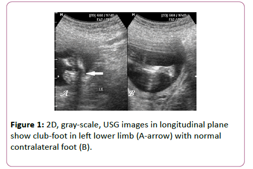
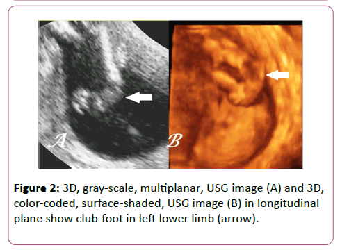
USG scan revealed a singleton pregnancy with a 17-18 week foetus showing positional abnormality of left foot with persistent adduction & inward rotation and normal liquor (Figure 1) without obvious abnormality in contralateral foot. Though the abnormality was suspected on 2D-mode yet 3D-mode played a key role in confirming the diagnosis (Figure 2). No evidence of any other sonographic fetal abnormality was noted. Based on the above findings, isolated, unilateral, club-foot was diagnosed.
The lady was counselled about the fetal anomaly with its inherent prognosis and was followed-up in postpartum period when clinical examination vaginally-delivered; full-term baby confirmed the antenatal diagnosis.
Discussion
Club-foot or Congenital Talipes Equinovarus (CTEV) is a common clinical condition with an estimated incidence of 1-3 per 1000 live births and male predominance [1,2]. It is a developmental structural abnormality of foot with hindfoot in equinus & varus position; midfoot in supination & forefoot in adduction. It is commonly associated with hypoplasia of ipsilateral leg as well. This condition needs differentiation from positional abnormality of foot as latter is fully correctable with positioning methods.
CTEV may occur in association with other conditions or as part of genetic syndromes (as arthrogryposis, trisomy 18, congenital myotonic dystrophy, neural tube defects, congenital cleft lip & palate, congenital heart disease, dysplastic disease of hip, amniotic band syndrome, etc.) in up to 10% of cases when it is considered complex else it may be considered as an isolated anomaly [1-3]. Oligohydramnios & multiple gestations have also been implicated as extrinsic factors in isolated cases [1]. The condition is usually unilateral but may be bilateral in up to one-third with bilateral cases being more severe than the unilateral ones [2,4]. Bilateral cases show similar involvement of both sides in terms of severity [4].
CTEV can be diagnosed prenatally by USG with variable degree of accuracy (maximum in second trimester) and false positive rates ranging from 0-40% with higher false-positivity in unilateral disease [1,2,5-7]. The major cause of false positive rate of CTEV is transient positioning of fetal foot which persists for less than 30 min making extended USG examination beyond half-an-hour imperative [7].
Until recently, the most commonly used criterion for diagnosis of CTEV on 2D-USG has been simultaneous visualisation of leg and foot bones in the same plane [6]. However, recent advent of 3D-USG has resulted in better in-vivo, morphological assessment of variety of fetal anomalies attributed to its multiplanar and surface-shaded modes [8].
Isolated unilateral CTEV is not an indication of karyotyping if a dedicated anomaly scan has been done especially with 3DUSG to rule out complex cases as it is not commonly associated with aneuploidy [1,2,6]. However in approximately 10-13% cases, the parents need to be counselled that prenatally-diagnosed, isolated CTEV may have complex CTEV detected in postnatal period and up to 10% may require minimal treatment.
Conclusion
3D-USG is a confident method of diagnosing isolated-CTEV which in general shows excellent postnatal outcome. Though a small risk of postnatal-detection of few anomalies is associated with isolated-CTEV yet amniocentesis is not routinely indicated especially if dedicated anomaly scan with 3D-USG has been performed.
References
- Lauson S, Alvarez Z, Patel MS, Langlois S (2010) Outcome of prenatally diagnosed isolated clubfoot. Ultrasound ObstetGynecol35: 708-714.
- Malone FD, Marino T, Biwanchi DW, Johnston KD, Alton ME (2000) Isolated Clubfoot Diagnosed Prenatally: Is Karyotyping Indicated? ObstetGynecol95: 437-440.
- Furdon SA, Donlon CR (2002) Examination of the newborn foot: positional and structural abnormalities. Adv Neonatal Care2: 248-258.
- Gray K, Barnes E, Gibbons P, little D, Burns J (2014) Unilateral versus bilateral clubfoot: an analysis of severity and correlation. J PediatrOrthop B 23: 397-399.
- Pagnotta G, Maffulli N, Aureli S, Maggi E, Mariani M, et al. (1996) Antenatal sonographic diagnosis of clubfoot: a six-year experience. J Foot Ankle Surg35: 67-71.
- Mammen L, Benson CB (2004) Outcome of foetuses with Clubfeet Diagnosed by Prenatal Sonography. J Ultrasound Med 23: 497-500.
- Keret D, Ezra E, Lokiec F, Hayek S, et al. (2002) Efficacy of Prenatal Ultrasonography in confirmed club-foot. J Bone Joint Surg Br 84:1015-1019.
- Khodair SA, Hassanen OA (2014) Abnormalities of fetal rib number and associated fetal anomalies using three-dimensional ultrasonography. The Egyptian Journal of Radiology and Nuclear Medicine 45: 689-694.
Open Access Journals
- Aquaculture & Veterinary Science
- Chemistry & Chemical Sciences
- Clinical Sciences
- Engineering
- General Science
- Genetics & Molecular Biology
- Health Care & Nursing
- Immunology & Microbiology
- Materials Science
- Mathematics & Physics
- Medical Sciences
- Neurology & Psychiatry
- Oncology & Cancer Science
- Pharmaceutical Sciences


