A Wrist Median Nerve Schwannoma: A Case Report
Zakariaa Ilias*
Department of Clinical Science, Hassan II University, Casablanca, Morocco
- *Corresponding Author:
- Zakariaa Ilias, Department of Clinical Science, Hassan II University, Casablanca, Morocco, Tel: 00212671247953; E-mail: zakariailias@gmail.com
Received date: March 07, 2022, Manuscript No. IPCRCR-22-12937; Editor assigned date: March 10, 2022, PreQC No. IPCRCR-22-12937 (PQ); Reviewed date: March 25, 2022, QC No. IPCRCR-22-12937; Revised date: May 09, 2 022, Manuscript No. IPCRCR-22-12937 (R); Published date: May 17, 2022, DOI: 10.36648/IPCRCR.6.5.001
Citation: Ilias Z (2022) A Wrist Median Nerve Schwannoma: A Case Report. J Clin Radiol Case Rep Vol:6 No:5
Abstract
Schwannoma’s are benign tumors that can affect all nerves even the peripheric nerve. We report a case of 66 year old man which had a mass upon his wrist with a diagnostic delay of two years. Diagnosis was based on imaging features (Ultrasonography and MRI) with Electromyography (EMG) which was assessed by histopathological examination.
Keywords
Schwannoma; Wrist; Median nerve; MRI
Introduction
The schwannoma’s are benign tumor which grows at the expense of schwann cells of the nerve sheath. The most common localizations are depending central nerve’s specially the cochleovestibular nerve. However it can be founded in peripheric nerves and it is regarded as the most common benign peripheral nerve tumor. When those tumors are small, they are usually painless and are responsible for diagnostic delay. However, when they are reaching a big size they can be responsible of paresthesias, pain and motor weakness. Imaging specially MRI and ultrasonography in addition of Electromyography (EMG) are the principal explorations for diagnosis providing a large set of informations that helps for the diagnosis. We report a case of a median nerve schwannoma in 68 years old patient who developed a wrist nodular mass which imaging have revealed a median nerve schwanomma.
Case Presentation
It’s a 66 years old man, who was working as an institutor and was a hypertensive patient which presented a nodular mass upon the ventral side of the wrist. This mass had appeared about 2 years; without any other sign such as parenthesis or motor weakness. This motivated our patient to consult a traumatologist. The clinical examination has shown a well circumbscribed mass upon the ventral side of the wrist, this mass was a soft mass attached to deeper tissues and the percussion over the mass provoked paresthesias along the path of median nerve (tinel like sensation). Those finding led the clinician to prescribe an ultrasonography and an EMG which has shown the following features.
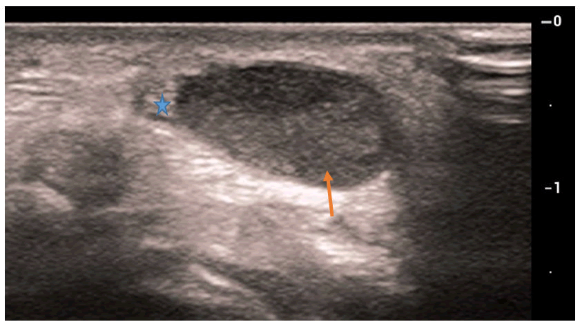
The ultrasonography has shown these following features (Figure 1):
- An ovalar well circumscribed hypoechoic mass depending the peripheric part of the median nerve measuring
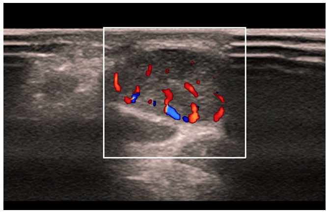
- This mass is well vascularized on Doppler color and Energy (Figure 2).
- This mass is located between FCR and palmary longus
- Without any visible communication with the tendons or the joint capsule
EMG has shown the following features:
- A conduction block depending the median nerve at the level of the Wrist estimated at 50% without any alteration of the motor distal latency or the motor conduction Velocity
- Whereas the parameters of the sensitive branches of the median nerve were completely normal
Following those examinations, a tumor depending the median nerve was truly suspected and an MRI of the Wrist was performed for better characterization following this protocol:
AT1 weighted sequences on sagittal and coronal planes
AT2 FATSAT sequences on sagittal axial and coronal planes
AT1 FATSAT sequences on sagittal axial and coronal planes
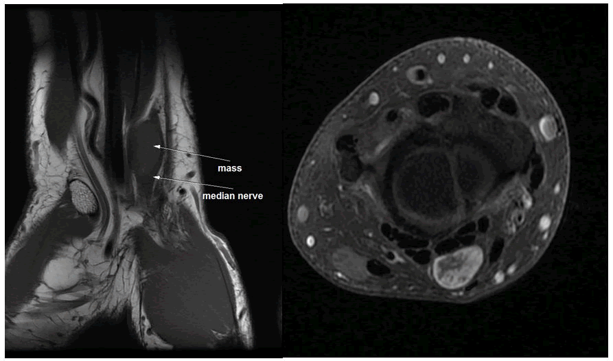
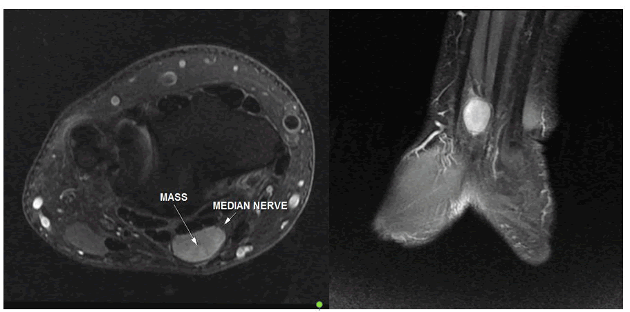
The MRI has found depending the palmar side of the wrist upon the superior part of the palmar tunnel an ovoid encapsulated mass measuring 14 x 19 x 7 mm. This mass has an intermediate signal on T1 weighted images; homogenous high signal in T2 weighted sequences. After Gadolinium administration this mass had a heterogenous enhancement. (Figures 3,4)
Furthermore, this mass is developed depending the peripheric part of the median nerve and seems to push back its nervous fibers.
Figure 3: MRI of the wrist in T1 weighted sequence in sagittal plan A) and on T1 FATSAT sequence after gadolinium administration in axial plan; B) showing an encapsulated mass with a heterogenous enhancement after gadolinium administration.
After all these examinations a surgery was performed which consists of a mass resection, the anatomopathological findings have confirmed the diagnosis by stating a median nerve schawannoma.
Discussion
Benign nerve sheath tumors are rare conditions in which there is an abnormal growth within the cells of this covering. Schwannoma’s and neurofibromas are the most common histological type [1]. Schwannomas are common, slowly growing, and encapsulated benign nerve sheath tumors separated from the surrounding tissues. Some forms may be localized within the nerve trunk or bundles of nerve fibers spreading over the surface of the tumor [2]. Epidemiologically, they are regarded as the most common peripheral nerve tumors in adults; they usually occur when the individual is between 30 and 50 years old, with no significant difference regarding ethnicity or gender [3]. The ulnar and median nerves are the most affected nerve [4-6]. There are several studies talking about median nerve schwannoma but only few are talking about its involvement on the wrist. The schwannoma is developed in the nerve sheath, and can occur anywhere within the peripheral nervous system. Most of the cases, this tumors are asymptomatic, and occasionally small palpable tumor mass, with few or no neurological deficit can be found [7]. It can produce pain, a positive Tinel’s sign or a Tinel’s-like sensation, and sensory alterations. The slow growth pattern of benign nerve tumors allows for adaptation of the nerve function to the pressure effects [8]. The slow growth allows a nervous adaptation which is responsible of the diagnostic delay. In our case the delay was about two years whereas A Sanoussi and Dubert have estimated an average period delay of nine month [9]. If ENMG can guide the clinician about median nerve damage especially in advanced tumors; imaging is the key for preoperative diagnosis and extension of those tumors. Although they do not have specificity, they are endowed with a certain diagnostic value, and some radiological characteristics can help doctors differentiate these tumors, helping to guide their approach [10]. On US, schwannomas are seen as well-defined, ovoid, heterogeneous masses most often in the peripheric part of the nerve [11]. The presence of blood flow on the Doppler can distinguish a peripheric nerve steam tumor from a cystic lesion. The MRI enables the characterization of lesions with neural involvement, their relation to important anatomical structures, and the extent of intrinsic or extrinsic nerve involvement. The lesion is seen as a well-defined mass in fusiform shape, located within the peripheric part of the nerve, is or hypointense in the signal related to the skeletal muscle in T1-weighted images, and with increased signal intensity and slightly heterogeneous in T2-weighted images Gadolinium enhanced MRI is useful in the diagnosis of schwannomas, with the characteristic “target sign”, that is a biphasic or triphasic pattern, in which the periphery has a higher intensity on the T2 sequence and low intensity on the Gadolinium enhanced T1 and vice versa on the central part. It was shown to have a specificity of 100% and a sensitivity of 59% in a histologically verified study by Koga et al [12]. Schwannomas and neurofibromas are not always easy to distinguish and radiological signs suggest that the former is in an eccentric position in relation to the parent nerve and heterogenous appearance. Calcifications can be seen in chronic lesions, the so called ancient schwannomas [13,14]. Surgical excision is regard to be the most effective method of therapy by many authors; however some authors prefer to excise only symptomatic tumours or those demonstrating enlargement during followup [15,16]. Careful microsurgical dissection is important by using a loupe or microscopical magnification in order to avoid the nerve fibers damages during the epineural and endoneurial dissection. Paresthesia is the most common postoperative complication [17]. The surgeon should be careful not to make unnecessary sacrifice of functionally important motor and sensory branches.
Conclusion
Schwannoma’s are benign tumor depending schwann cells of the nerve sheath. If they are most frequently depending the central nerves they can also affect peripheric nerve and are regarded as the most frequent histologic type of peripheric nerve tumors. This tumors are often asymptomatic responsible for diagnostic delay. Imaging specially MRI represents the gold standard for the diagnostic providing a large set of informations for the diagnosis.
References
- Shanouda S, Kaya G (2017) Benign cutaneous peripheral nerve sheath tumor with hybrid features: report of two cases with schwannoma/perineurioma and schwannoma/neuro fibroma components. Dermatopathol 4:1-6
[Crossref] [Google Scholar] [Indexed]
- Boufettal M, Azouz M, Rhanim A, Abouzahir M, Mahfoud M, et al. (2014) Schwannoma of the median nerve: diagnosis sometimes delayed. Clin Med Insights Case Rep 7:71-73
[Crossref] [Google Scholar] [Indexed]
- Magalhaes MJS, Perreira DVM, Oliva HNP, Duraes DTS, Caiado FL, et al. (2019) Peripheral nerve schwannomas: A literature review. Arq Bras Neurocir 38:308–314
[Crossref] [Google Scholar] [Indexed]
- Patel MR, Mody K, Moradia VJJ (1996) Multiple schwannomas of the ulnar nerve: a case report. Hand Surg Am 21:875-876
[Crossref] [Google Scholar] [Indexed]
- Kang HJ, Shin SJ, Kang ES (2000) Schwannomas of the upper extremity. J Hand Surg Br 25:604-607
[Crossref] [Google Scholar] [Indexed]
- Kutahya H, Guleç A, Guzel Y, Kacira B, Toker S (2013) Schwannoma of the median nerve at the wrist and palmar regions of the hand: a rare case report. Case Rep Orthop 2013:950106
[Crossref] [Google Scholar] [Indexed]
- Mackinnon SE (2015) Nerve Surgery. Thieme Medical Publishers, New York.
- Ogose A, Hotta T, Morita T, Yamamura S, Hosaka N, et al. (1999) Tumors of peripheral nerves: correlation of symptoms, clinical signs, imaging features, and histologic diagnosis. Skeletal Radiol 28:183-188
[Crossref] [Google Scholar] [Indexed]
- Akambi Sanoussi K, Dubert T (2006) Schwannomas of the peripheral nerve in the hand and the upper limb: Analysis of 14 cases. Chir Main 25:131-135
[Crossref] [Google Scholar] [Indexed]
- Arabi H, Khalfaoui S, El Bouchti I, Rokhsi R, Benhima MA, et al. (2015) Meralgia paresthetica with lumbar neurinoma: Case report. Ann Phys Rehabil Med 58:359–361
[Crossref] [Google Scholar] [Indexed]
- Madi S, Pandey V, Mannava K, Acharya K (2016) A benign ancient schwannoma of the tibia nerve. BMJ Case Rep 2016:1–2
[Crossref] [Google Scholar] [Indexed]
- Koga H, Matsumoto S, Manabe J, Tanizawa T, Kawaguchi N (2007) Definition of the target sign and its use for the diagnosis of schwannomas. Clin Orthop Relat Res 464:224-229
[Crossref] [Google Scholar] [Indexed]
- Lin J, Martel W (2001) Cross-sectional imaging of peripheral nerve sheath tumors: characteristic signs on CT, MR imaging, and sonography. AJR Am J Roentgenol 176:75-82
[Crossref] [Google Scholar] [Indexed]
- Harkin JC (1968) Tumors of the peripheral nervous system. Armed Forces Institute of Pathology, Washington, Dc, USA.
- Gonzalvo A, Fowler A, Cook RJ (2011) Schwannomatosis, sporadic schwannomatosis, and familial schwannomatosis: a surgical series with long-term follow-up: clinical article. J Neurosurg 114:756–762
[Crossref] [Google Scholar] [Indexed]
- Aslam N, Kerr G (2003) Multiple schwannomas of the median nerve: a case report and literature review. Hand Surg 8:249–252
[Crossref] [Google Scholar] [Indexed]
- Lee SH, Jung HG, Park YC, Kim H-S (2001) Results of neurilemona treatment: a review of 78 cases. Orthopedics 24:977–980
[Crossref] [Google Scholar] [Indexed]
Open Access Journals
- Aquaculture & Veterinary Science
- Chemistry & Chemical Sciences
- Clinical Sciences
- Engineering
- General Science
- Genetics & Molecular Biology
- Health Care & Nursing
- Immunology & Microbiology
- Materials Science
- Mathematics & Physics
- Medical Sciences
- Neurology & Psychiatry
- Oncology & Cancer Science
- Pharmaceutical Sciences
