ISSN : ISSN: 2576-1455
Journal of Heart and Cardiovascular Research
A Case of Pericardial Metastasis: Image Case Report
Mahmoud H Abdelnaby1, Abdallah M Almaghraby2 and Ashraf A El-Amin2
1Cardiology and Angiology Unit, Department of Clinical and Experimental Internal Medicine, Medical Research Institute, University of Alexandria, Alexandria, Egypt
2Department of Cardiology, University of Alexandria, Alexandria, Egypt
- *Corresponding Author:
- Mahmoud H Abdelnaby
Cardiology and Angiology Unit
Department of Clinical and Experimental Internal Medicine
Medical Research Institute, University of Alexandria, Egypt
Tel: +201007573530
E-mail: Mahmoud.hassan.abdelnabi@outlook.com
Received Date: 22 January 2018; Accepted Date: 06 February 2018; Published Date: 15 February 2018
Citation: Abdelnaby MH, Almaghraby AM, El-Amin AA (2018) A Case of Pericardial Metastasis: Image Case Report. J Heart Cardiovasc Res. Vol.2 No.1:1
Copyright: © 2018 Abdelnaby MH, et al. This is an open-access article distributed under the terms of the creative Commons attribution License, which permits unrestricted use, distribution and reproduction in any medium, provided the original author and source are credited.
Abstract
A 76-year-old male with a past history of resected cancer colon and a recurrence in the form of hepatic and lung metastases had a Chest X Ray (CXR) which revealed cardiomegaly and referred for echocardiography that revealed a large pericardial mass with pericardial effusion most likely metastatic in nature.
Keywords
Pericardium; Metastasis; Cancer colon
Introduction
Although primary cardiac tumors are extremely rare, secondary tumors are not. Theoretically the heart can be metastasized by any malignant neoplasm able to spread to distant sites.
Case Presentation
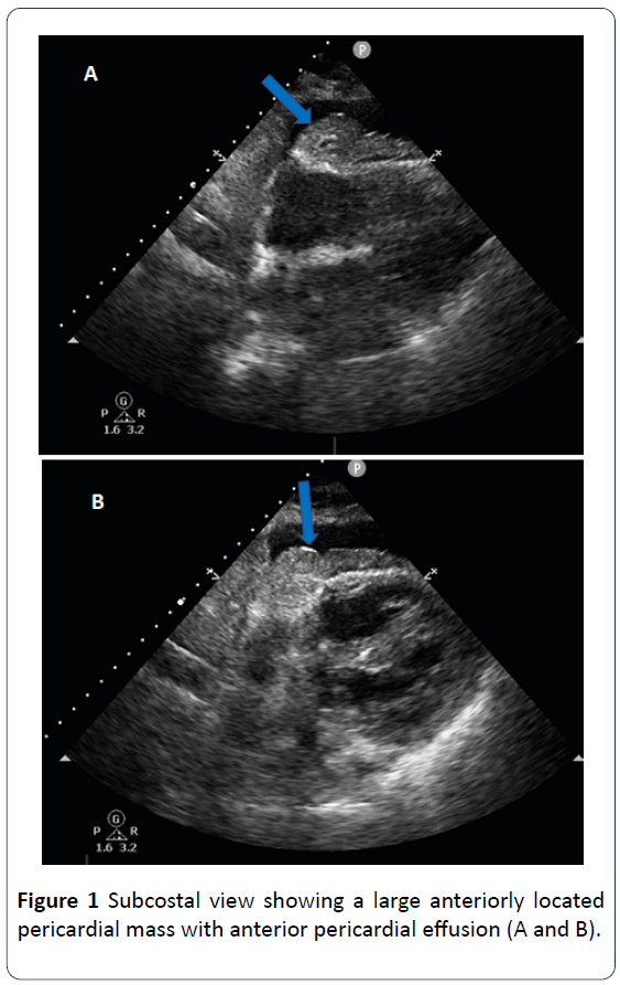
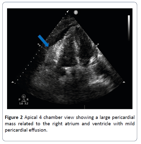
A 76-year-old male patient with past medical history of diabetes, hypertension, ischemic heart disease and chronic renal failure on maintenance hemodialysis and a history of resected adenocarcinoma of the colon 2 years ago with recurrence diagnosed 6 months ago in the form of hepatic and pulmonary metastases had a CXR which revealed cardiomegaly and bilateral pulmonary infiltrates. He was referred for echocardiography which showed a large fleshy pericardial mass attached to the anterior surface of the right atrium and ventricle with anterior pericardial effusion most likely metastatic in nature. Due to his critical condition, no further investigations were done and only supportive and palliative measures were adopted (Figures 1 and 2).
Discussion and Conclusion
Primary pericardial malignancies are extremely rare [1]. Metastases to the heart and pericardium are much more common than primary cardiac tumors and are generally associated with a poor prognosis [2]. Echocardiography remains the key method for diagnosis of cardiac masses [3]. Advances in other imaging modalities such as Cardiac Magnetic Resonance (CMR) and Cardiac Computed Tomography (CT) are associated with improved tissue characterization with better spatial and temporal resolutions [4].
References
- Restrepo CS, Vargas D, Ocazionez D, Martínez-Jiménez S, Betancourt Cuellar SL, et al. (2016) Primary pericardial tumors. Radiographics 33: 1613-30.
- Hoffmeier A, Sindermann JR, Scheld HH, Martens S (2014) Cardiac tumors-diagnosis and surgical treatment. Deutsches Ärzteblatt international 111: 205.
- Mankad R, Herrmann J (2016) Cardiac tumors: Echo assessment. Echo research and practice 3: R65-R77.
- Motwani M, Kidambi A, Herzog BA, Uddin A, Greenwood JP, et al. (2013) MR imaging of cardiac tumors and masses: A review of methods and clinical applications. Radiology 268: 26-43.
Open Access Journals
- Aquaculture & Veterinary Science
- Chemistry & Chemical Sciences
- Clinical Sciences
- Engineering
- General Science
- Genetics & Molecular Biology
- Health Care & Nursing
- Immunology & Microbiology
- Materials Science
- Mathematics & Physics
- Medical Sciences
- Neurology & Psychiatry
- Oncology & Cancer Science
- Pharmaceutical Sciences


