Volumetric Changes of Edentulous Jaws Beneath Conventional Complete Dentures
Mohamed Samih Alsrouji* and Rohana Ahmad
Department of Prosthodontics, Faculty of Dentistry, University Technology Mara, Malaysia
- Corresponding Author:
- Alsrouji MS
Department of Prosthodontics
Faculty of Dentistry
University Technology Mara, Malaysia
Tel: 0060176821075
E-mail: alsrouji@yahoo.com
Received Date: June 13, 2017; Accepted Date: July 05, 2017; Published Date: July 09, 2017
Citation: Alsrouji MS (2017) Volumetric Changes of Edentulous Jaws Beneath Conventional Complete Dentures. Pros Orth Open J 1:10.
Copyright: © 2017 Alsrouji MS. This is an open-access article distributed under the terms of the Creative Commons Attribution License, which permits unrestricted use, distribution, and reproduction in any medium, provided the original author and source are credited.
Abstract
Objective: The objective of this study was to quantify the changing in edentulous maxilla and mandible bone beneath complete dentures one year post dentures insertion and to create a visual map of these changes.
Materials and Methods: Four edentulous patients were provided by conventional complete dentures. CBCT were acquired for these patients prior and one year post dentures insertion. These radiographs converted to Mimics research software where a 3-D model for maxilla & mandible were calculated pre and post treatment for each patient. For each patient, the pre & post model for both jaws underwent to superimposition. Then these superimposed models were sectioned at the same level to ensure the same borders of jaws pre & post treatment. The bone volume pre & post treatment for both jaws obtained and the difference between these values calculated. Finally these models exported to 3-Matic software to create visualized maps for these changes.
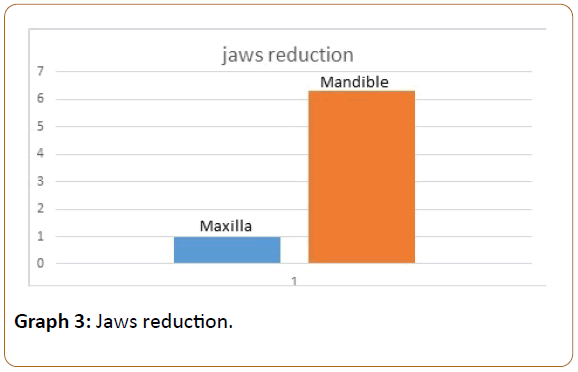
Results: The average of reduction in the maxilla was 0.97% while this average in mandible was 6.31%.
Conclusion: The three dimensional changes in the mandible one year post denture insertion was about six times more than maxilla. The high rate of resorption in mandible may complicate the mandible complete denture treatment so alternative treatment option should be considered for mandible such as implant overdenture.
Keywords
Maxilla and Mandible bone; CBCT; Dentures; Radiographs; Superimposition; Resorption; Overdenture
Introduction
Conventional complete dentures have been used to restore the edentulous patients for many decades. Several studies showed an adequate patient’s satisfaction with complete dentures. Carlsson reported 80 to 90% of patient’s satisfaction with complete dentures [1]. Ellis found significant improvement in patient’s satisfaction and life’s quality either in conventional or duplicate denture [2]. Regarding complete dentures, Santos showed that the patient’s satisfaction was more than expectation in term of chewing, esthetics, and phonetic [3].
In the last three decades dental implants have been used widely to stabilize the dentures. Mandible overdenture has been recommended as the minimal standard of care for edentulous patients [4]. Studies showed superiority of overdenture in psycho-social [5], nutrition [6], and function [7] issues. Despite these advantages of implant-overdenture, conventional complete denture still widely used to restore edentulous patients. Number of edentulous patient in USA only expects to be 38 million in 2020 due to increasing age of the general population [8]. The prevalence of edentulism in other countries varied between 3- 80% among the elderly over 60 [9]. Large number of these people is not interested in implant overdenture and they prefer complete denture due to economic or medical conditions [10].
Bone resorption under complete dentures is one the most drawbacks of complete dentures, particularly mandibular denture. Depending on cephalometric radiographs, Atwood measured 0.1 mm and 0.4 mm annual bone loss in anterior maxillary and mandibular ridge respectively, the mandibular ridge loss was 4 times greater than maxillary ridge [11]. In 7 years period of follow up post dentures insertion, Tallgren also found resorption in mandibular ridge 4 times than maxilla one [12].
Al the previous studies utilized 2-D radiographs. The objective of this study was to quantify the maxillary and mandibular bone loss 1-year post denture insertion in 3-D manner to measure the resorption more precisely and compare these changes with other studies that used 2-D manner, as well as to create a clear visualized map of this resorption in both jaws.
Materials and methodology
Four full edentulous patients were recruited for this study. All the patients were healthy without serious diseases, American Society Anesthesiologists classification 1 and 2 (ASA 1 and 2). They restored with conventional complete dentures in balance occlusion. All the prosthetic works in the clinic and the laboratory were done by one clinician and one technologist.
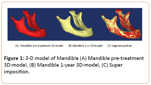
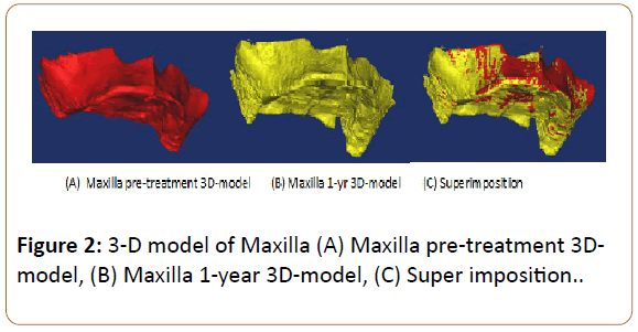
CBCT radiographs were taken at two stages, the first prior to treatment and the second one year post treatment. Also these images were taken by one radiographic technologist and by utilizing the same CBCT machine and parameters (i-CAT.120 kVp, 18.45 mAs, and 20-s). These images were converted to Mimics research software and underwent to cautious segmentation, subsequently a 3-D model of mandible and maxilla were calculated pre and 1 yr post treatment (Figures 1 and 2). The models were first subjected to manual superimposition by closing the models as possible then superimposition was finalized using the Standard Tessellation Language technique (STL).
Figure 1: 3-D model of Mandible (A) Mandible pre-treatment 3D-model, (B) Mandible 1-year 3D-model, (C) Super imposition.
Figure 2: 3-D model of Maxilla (A) Maxilla pre-treatment 3Dmodel, (B) Maxilla 1-year 3D-model, (C) Super imposition.
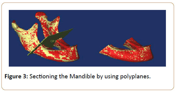
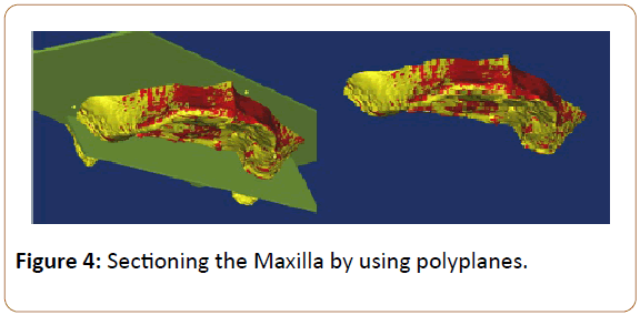
Following that, polyplanes were used to section the required parts from both jaws which contain the bearing areas and supporting bone and eliminate the other structures as well as to ensure the same borders of pre & post models (Figures 3 and 4).
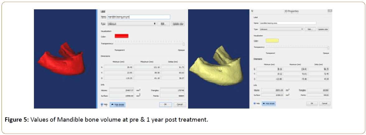
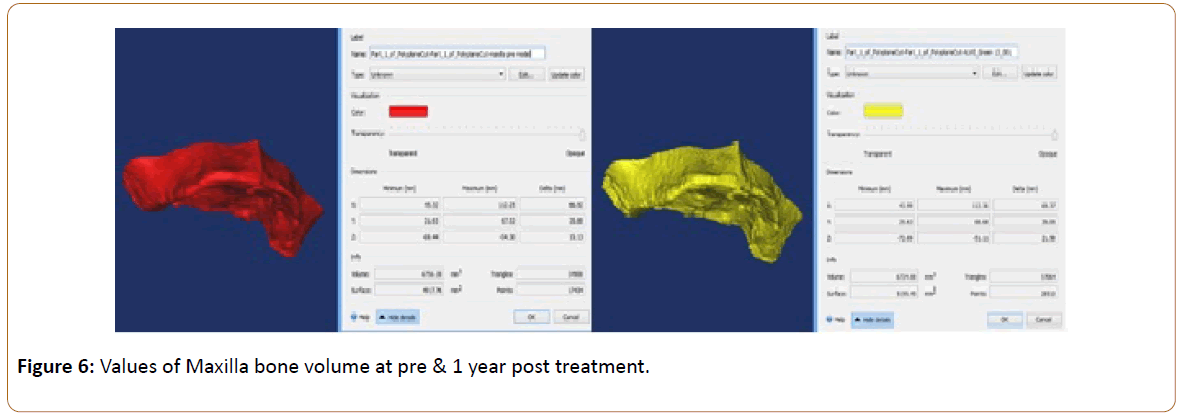
The bone volume values were obtained from the software for pre & post model of both jaws and the percentages of changing were calculated for each patient (Figures 5 and 6).
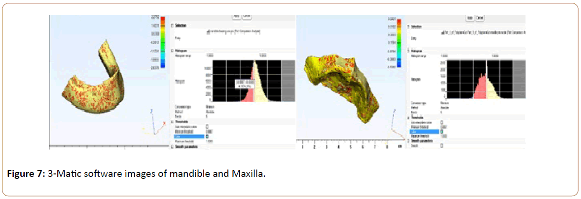
Finally these models were sent to 3-Matic software to create a clear visualized map of region and depth of jaws changing (Figure 7).
Results
The results of mandible and maxilla bone changing arranged in Table 1 and 2 respectively (Tables 1 and 2).
| Volume of Mandible at Pre-treatment (mm3) | Volume of Mandible at 1 year Post-treatment (mm3) | Mandible Volume Change (%) | |
|---|---|---|---|
| 1. | 29478.37 | 28091.08 | -4.71 |
| 2. | 37451.06 | 33977.18 | -9.28 |
| 3. | 22218.45 | 20931.32 | -5.79 |
| 4. | 27812.9 | 26295.73 | -5.45 |
| Average | -6.31 |
Table 1: Change in the mandible bone volume.
| Volume of Maxilla at Pre-treatment (mm3) | Volume of Maxilla at 1 year Post-treatment (mm3) | Maxillary Volume Change (%) | |
|---|---|---|---|
| 1. | 6756.28 | 6724.88 | -0.46 |
| 2. | 10736.94 | 10694.35 | -0.40 |
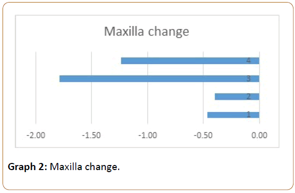
| 3. | 5879.45 | 5774.28 | -1.79 |
| 4. | 7897.23 | 7799.78 | -1.23 |
| Average | -0.97 |
Table 2: Change in the maxilla bone volume.
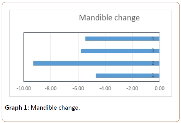
For the mandible changing the mean was–6.31%, standard deviation SD=1.02%, median=-5.62%, and range=-9.28% to– 4.71% (Graph 1).
While in maxilla the mean was –0.97%, standard deviation SD=0.66%, median=0.85%, and range=-1.79% to–0.40% (Graph 2).
Discussion
Despite high use of dental implants in restore dental function in edentulous patients over the last few decades, conventional complete dentures still the main method of restoring those patients [13]. Complete dentures able to restore aesthetics, function, facial contour, and satisfaction [14,15]. The continuous resorption of mandible residual ridge may contribute to retention loss of mandible denture with time. Bone resorption following tooth extraction is a continuous permanent process. Several reasons contribute to this phenomenon: anatomical, metabolic, functional, and prosthetic reasons [16]. Several studies tried to measure the bone reduction under dentures by utilizing 2-D radiographs. Atwood and Tallegren found that bone resorption in mandible is four times more than maxilla [11,12]. While in this 3-D study the mandible resorption was six times more than maxilla.
The volume of reduction in mandible in this study appears to be higher than known from the previous studies while it is almost the same for maxilla (Graph 3). This high rate of mandible resorption will certainly reduce the prognosis of mandible denture as well as encourages patients and clinicians to use implant-retained overdenture to restore mandible.
Conclusion
Bone resorption associated with conventional complete denture is much higher in mandible than maxilla as previous studies confirmed. In this 3-D study the rate of this reduction was about 6:1 (mandible to maxilla) and it was greater than other showed in other 2-D studies which was 4:1. The high rate of bone resorption in mandible affects the mandible denture retention. As a large number of patients still prefer complete dentures rather implant overdenture then more investigations are required to avoid or minimize this resorption. Furthermore, patient education regarding the importance and effectiveness of implant-retained overdenture is required.
References
- Carlsson GE (2006) Facts and fallacies: An evidence base for complete dentures. Dent Update 33: 134-142.
- Ellis JS, Pelekis ND, Thomason JM (2007) Conventional rehabilitation of edentulous patients: The impact on oral health-related quality of life and patient satisfaction. J Prosthodont 16: 37-42.
- Santos BFO, dos Santos MB, Santos JF, Marchini L (2015) Patients' evaluations of complete denture therapy and their association with related variables: A pilot study. J Prosthodont 24: 351-357.
- Feine JS, Carlsson GE, Awad MA, Chehade A, Duncan WJ, et al. (2002) The McGill consensus statement on overdentures. Montreal, quebec, Canada. May 24-25, 2002. Int J Prosthodont 15: 413-414.
- Heydecke G, Thomason JM, Lund JP, Feine JS (2005) The impact of conventional and implant supported prostheses on social and sexual activities in edentulous adults Results from a randomized trial 2 months after treatment. J Dent 33: 649-657.
- Morais JA, Heydecke G, Pawliuk J, Lund JP, Feine JS (2003) The effects of mandibular two-implant overdentures on nutrition in elderly edentulous individuals. J Dent Res 82: 53-58.
- Van der Bilt A, Burgers M, Van Kampen FM, Cune MS (2010) Mandibular implant-supported overdentures and oral function. Clin Oral Implants Res 21: 1209-1213.
- Douglass CW, Shih A, Ostry L (2002) Will there be a need for complete dentures in the United States in 2020? J Prosthet Dent 87: 5-8.
- Nitschke N (2004) To the oral health of seniors. Habilitation, University of Leipzig, Leipzig.
- Felton DA (2009) Edentulism and comorbid factors. J Prosthodont 18: 88-96.
- Atwood D, Coy W (1971) Clinical, cephalometric, and densitometric study of reduction of residual ridges. J Prosthet Dent 26:280-295.
- Tallgren A (1970) Alveolar bone loss in denture wearers as related to facial morphology. ActaOdontolScand 28: 251-270.
- Carlsson GE, Omar R (2010) The future of complete dentures in oral rehabilitation. A critical review. J Oral Rehabil 37: 143-156.
- Berg E (1993) Acceptance of full dentures. Int Dent J 43: 299-306.
- Bellini D, Dos Santos MB, De Paula Prisco DCV, Marchini L (2009) Patients' expectations and satisfaction of complete denture therapy and correlation with locus of control. J Oral Rehabil 36: 682-686.
- Atwood DA (1971) Reduction of residual ridges: A major oral disease entity. J Prosthet Dent 26: 266-279.

Open Access Journals
- Aquaculture & Veterinary Science
- Chemistry & Chemical Sciences
- Clinical Sciences
- Engineering
- General Science
- Genetics & Molecular Biology
- Health Care & Nursing
- Immunology & Microbiology
- Materials Science
- Mathematics & Physics
- Medical Sciences
- Neurology & Psychiatry
- Oncology & Cancer Science
- Pharmaceutical Sciences
